Skip to content
Prof. Dr. Ali Avanoğlu
Main treatment page • UPJ Obstruction

UPJ Obstruction Surgery in Turkey

UPJ obstruction (ureteropelvic junction obstruction; informally, kidney outlet narrowing) is a slowing of urine drainage from the kidney caused by a narrowing at the point where the renal pelvis opens into the ureter. In infants and children, it's most often noticed while investigating hydronephrosis (kidney swelling) seen on a prenatal or post-birth ultrasound; but not every case of hydronephrosis is UPJ obstruction, and a significant share resolves on its own. Diagnosis starts with ultrasound and is completed with MAG3 diuretic renography (a kidney scan), which measures the kidney's share of function and how fast it drains. Treatment is not a single path: a well-functioning, stable kidney is followed with serial ultrasound and, if needed, repeat MAG3; if function is dropping, widening is increasing, or there are symptoms, pyeloplasty (repair of the narrowing) comes onto the table. This page walks through that whole picture.

ByProf. Dr. Ali AvanoğluUpdated
At a glance 45 pages • 3 video
~1–5% of pregnancies
Prenatal hydronephrosis

Mostly mild and temporary; UPJ obstruction is the most common pathological cause

40% threshold
Split function

Good function + good drainage + stable ultrasound → usually follow-up

~90–95% success
Pyeloplasty

Anderson–Hynes repair; open, laparoscopic, or robotic

The whole treatment • 12 sections

This page explains the whole picture; the details of each topic are in the pages below

Start with what UPJ obstruction is and how it differs from hydronephrosis; then its causes, symptoms, the path to diagnosis, the degree of hydronephrosis, MAG3 and function, the follow-up-or-surgery decision, conservative follow-up, pyeloplasty, the open–laparoscopic–robotic approaches, what comes after surgery, and special situations: twelve sections. Each section has detail pages below it; jump to any section from the table of contents on the right (at the top on mobile).

what is kidney enlargement
01Section 1 / 12

What is UPJ obstruction (ureteropelvic junction obstruction, kidney outlet narrowing)? How it differs from hydronephrosis

A narrowing at the kidney's outlet slows urine flow; hydronephrosis is a finding, not a diagnosis

The kidney produces urine; urine first collects in small chambers inside the kidney (the calyces), then gathers in a funnel-shaped chamber, the renal pelvis, and from there travels down a thin channel called the ureter to the bladder. The point where the pelvis opens into the ureter is called the ureteropelvic junction (UPJ; sometimes written PUJ, or "UP junction," in reports). If the passage at this point is narrow or unable to contract, urine backs up, and the pelvis and calyces widen. This is where the everyday name "kidney outlet narrowing" comes from.

RIGHTLEFT
SFU 3 hydronephrosis on one side: pelvis wide, narrowing at the UPJ (red ring), ureter normal; other side normal
Definitionthree sentences
  • 1A narrowing at the kidney's outletat the point where the renal pelvis opens into the ureter, urine flow slows; the pelvis and calyces widen
  • 2Hydronephrosis is a finding, UPJ obstruction is a diagnosisnot every widened kidney means a narrowing; the most common cause of prenatal hydronephrosis is temporary widening
  • 3Most are followed without surgeryroughly a quarter of children with UPJ-type hydronephrosis eventually need pyeloplasty; improvement mostly happens in the first 2–3 years
Quick factsat a glance
Frequency~1/1,000–1,500 live births; ~2× more common in boys; more often on the left; bilateral in ~10–30% of newborns
DiagnosisUltrasound (AP diameter, SFU/UTD) + MAG3 diuretic renography; VCUG in selected cases
CourseMild hydronephrosis mostly improves; ~20–30% pyeloplasty at SFU 3–4
TreatmentConservative follow-up or pyeloplasty (open / laparoscopic / robotic)
TeamDecision and surgery by two specialists; follow-up continues into adulthood
Hydronephrosis ≠ UPJ obstruction

Prenatal hydronephrosis occurs in ~1–5% of pregnancies; ~40–80% is temporary/physiological. UPJ obstruction (~10–30%) is the most common pathological cause; VUR, a lower ureteral narrowing, PUV and a duplex system are the others.

Read the full text
  • UPJ obstruction occurs in roughly 1 in 1,000–1,500 live births; it is about twice as common in boys as in girls, and more often seen on the left side. Both kidneys being affected together in the newborn period has been reported in the literature at roughly 10–30%. The large majority of cases are congenital; now that prenatal ultrasound is widespread, most infants are noticed before any symptom appears, either before birth or within the first weeks after.
  • One distinction needs to be made from the start: hydronephrosis is widening of the kidney's collecting system with urine — it is a finding, not a diagnosis. Widening has many causes: temporary (physiological) widening is the most common; UPJ obstruction, kidney reflux (vesicoureteral reflux, VUR), narrowing where the ureter enters the bladder (megaureter), posterior urethral valves in boys (PUV), and a duplex collecting system are others. UPJ obstruction is the most common pathological cause of newborn hydronephrosis, but the single most common cause overall is temporary widening. So the sentence "there's hydronephrosis on the ultrasound" does not mean "there's a narrowing in the kidney."
  • If the cause of the hydronephrosis turns out to be kidney reflux, the path diverges. This page and the pages beneath it are about children whose widening is caused by — or suspected to be caused by — narrowing at the UPJ level.
  • The significance of UPJ obstruction is that kidney tissue under prolonged pressure can lose function over time. But in most children the narrowing is partial; the kidney adapts as it grows and keeps its function. So the real question is not "is there a narrowing" but "is this kidney being harmed" — and the answer comes not from a single test but from the course followed with MAG3 and serial ultrasound.
Hydronephrosis is a finding, not a diagnosis

The word "hydronephrosis" on an ultrasound report tells you the kidney is widened — it doesn't tell you why. Most of the widening found before birth is temporary and resolves on its own; UPJ obstruction accounts for a share of these cases. Separating the cause requires a repeat ultrasound and, if needed, MAG3.

Videos in this sectionFull guide (3) →
why does kidney swelling happen
02Section 2 / 12

Why does UPJ obstruction happen? Congenital narrowing, a crossing vessel, high ureteral insertion

Mostly a congenital narrow segment; a crossing vessel in older children

In children, UPJ obstruction is almost always congenital; it has nothing to do with anything done or not done during pregnancy, feeding, or infant care. Three structural types underlie the narrowing, and more than one can be present together in the same child. Which type predominates affects less the treatment method itself and more how the operation is planned.

Intrinsic narrow segmentthick wallnarrow lumenWALL MUSCLE / COLLAGEN DEFECT
Crossing vessellower pole vesselEXTERNAL COMPRESSION · INTERMITTENT BLOCKAGE
High ureteral insertionnarrow angleDOWNWARD FLOW DIFFICULT · KINK/FOLD
Three causes: intrinsic narrow segment, a crossing vessel to the lower pole, high ureteral insertion
Two main mechanismsinternal / external
Intrinsic narrowingthe junction's own wall
  • A narrow or non-contracting (aperistaltic) segment
  • Congenital; the main cause in infants
  • May be accompanied by a high insertion of the ureter into the pelvis
External compressiona crossing vessel
  • An extra vessel to the lower pole compresses the junction from outside
  • ~20–40% of childhood UPJ obstruction; approaching half in older children/adolescents with episodic pain
  • Dismembered pyeloplasty preserves the vessel, moving the ureter in front of it
How does the narrowing affect the kidney?three steps
  1. 1Urine collects in the pelvisoutflow is slow; the pelvis and calyces widen (hydronephrosis)
  2. 2Pressure reaches the parenchymaif prolonged, kidney tissue can thin and the function share can drop
  3. 3Stagnation can cause problemsit makes infection and stone formation easier
Read the full text
  • The most common type is intrinsic narrowing: a short segment of the junction's own wall is narrow or unable to contract (aperistaltic). In this segment the smooth muscle fibers are disorganized and connective tissue is increased; the contraction wave that carries urine down the ureter cannot pass through it. From the outside the ureter can look open, but functionally it does not let urine through. This is the main cause in infants.
  • The second type is a crossing vessel: an extra (accessory) artery or vein running to the kidney's lower pole passes in front of the junction and compresses it from outside. It is found in roughly 20–40% of childhood UPJ obstruction; in older children and adolescents, especially those presenting with episodic pain, the reported figure approaches half. The vessel is often not solely responsible — it frequently occurs together with intrinsic narrowing. During surgery, the vessel is preserved and the ureter is moved in front of it.
  • The third type is high ureteral insertion: the ureter leaves not from the lowest point of the pelvis but from its side or above. As the pelvis widens, this insertion point develops a kink and a valve-like effect, making emptying even harder. It is usually seen together with a noticeably widened pelvis; dismembered pyeloplasty corrects this as well, by moving the ureter to the lowest point of the pelvis.
  • Rarer or acquired causes also exist: a stone, scarring after previous surgery or infection, and association with kidney-shape anomalies such as a horseshoe kidney. These come up more often in adolescents and adults. When a crossing vessel is suspected, MR urography can give a clue; but which type predominates usually only becomes clear during surgery itself.
Was something done wrong?

No. UPJ obstruction is a structural difference that arises during the development of the kidney and ureter before birth. No link has been shown to diet, medication, or lifestyle during pregnancy; there is no way a family could have prevented it.

Prof. Dr. Ali Avanoğlu's publications on this topic · 1
  1. Avanoglu A, Tiryaki S (2020). Embryology and Morphological (Mal)Development of UPJ. Frontiers in pediatrics. PubMed ↗
kidney swelling symptoms
03Section 3 / 12

Symptoms of UPJ obstruction: silent in infants, episodic pain in older children (Dietl's crisis)

Silent in infants; episodic flank pain with vomiting in older children

In infants, UPJ obstruction usually causes no symptoms at all; the diagnosis is made from hydronephrosis seen on a prenatal or post-birth ultrasound. Less often, a febrile urinary tract infection, a soft mass felt in the abdomen (a very widened pelvis), unexplained fussiness, or vomiting and poor weight gain can be the first clue. This is why, in infants, the decision rests on ultrasound and MAG3 findings rather than symptoms.

RIGHTLEFT
Wide pelvis and a flank pain wave: stretching pain as urine builds up
How is it noticed?four paths
  • 1Hydronephrosis before birththe most common route to diagnosis; ultrasound ≥48 hours after birth, repeated at 4–6 weeks
  • 2Episodic flank pain + vomitingDietl's crisis: attacks after heavy fluid intake; in older children/adolescents; ultrasound during the pain matters
  • 3Febrile urinary tract infectioninfection in an obstructed kidney; same-day evaluation
  • 4A stone, blood in the urine, an incidental findingmore common in adolescents and adults
Read the full text
  • The picture changes in older children and adolescents: the most typical symptom is flank or abdominal pain. Its classic form is Dietl's crisis: sudden, severe flank or abdominal pain, nausea and vomiting after heavy fluid intake or something that increases urine output (drinking a lot of water, a watery meal, sometimes vigorous activity); the attack lasts hours and resolves on its own, and the child is completely well between episodes. Because the widened pelvis can shrink between attacks, an ultrasound taken at that time can even look normal — which is why an ultrasound taken during the pain itself is valuable for diagnosis. These attacks are often mistaken for appendicitis or a stomach bug.
  • Other symptoms are less common: a febrile urinary tract infection (an obstructed, infected kidney is an emergency), blood in the urine following minor trauma to a widened kidney, stone formation in stagnant urine, and, in a young child who can't localize it, recurring "stomach ache." A child may point to the belly-button area rather than the flank, which can delay diagnosis.
  • Hydronephrosis found before birth is not a symptom but a finding, and follows a separate path. In pregnancy, a pelvis AP diameter of 4 mm or more at 16–27 weeks, or 7 mm or more after 28 weeks, is placed under monitoring. After birth, the first ultrasound is done after at least 48 hours have passed, usually on day 3–7, because a newborn's relatively low fluid intake in the very first days can mask the widening. If the first ultrasound is normal or mild, it's repeated at 4–6 weeks; imaging is not delayed if there is severe bilateral hydronephrosis, a solitary kidney, low amniotic fluid in pregnancy, or suspected PUV.
See a doctor the same day

Fever (38 °C/100.4 °F and above) with flank or abdominal pain, or fever in a child with known hydronephrosis; severe, unrelenting flank or abdominal pain with vomiting; a drop in urine output in a solitary kidney or bilateral hydronephrosis. These can mean an obstructed, infected kidney or an acute obstruction.

Videos in this sectionFull guide (3) →
kidney swelling in a baby boy
04Section 4 / 12

How is it diagnosed? Ultrasound, when VCUG is needed, MAG3, MR urography

Ultrasound shows it; MAG3 measures function and drainage; VCUG in selected cases

Diagnosis proceeds in steps, and each step seeks an answer to two questions: what is causing the widening, and is the kidney being harmed? The usual order is ultrasound, VCUG if needed, then MAG3, and MR urography in selected cases. Not every child needs every test; which one is requested, and when, depends on the picture on ultrasound.

UltrasoundUreter dilated?Bladder abnormal? UTI?VCUGMAG3≥4–6 weeksMR urographyDecisionyesnoanatomy unclear?anatomy clear → decisionAfter birth ≥48 hours; AP diameter + SFUMAG3: waiting 4–6 weeks in infants for kidney maturation.VCUG: if there is ureteral dilation, a bladder abnormality, or febrile UTI.
Diagnostic pathway: ultrasound → VCUG if needed → MAG3 (4–6 weeks) → MR urography if anatomy unclear → decision
Testswhat, why, when
TestWhat it showsWhen it's used
UltrasoundAP diameter, calyx widening, parenchyma thickness, the ureter and bladderFirst test and follow-up; ≥48 hours after birth, repeated at 4–6 weeks
VCUG (catheter study)Whether there's reflux; whether the urethra is normal (PUV)Selected cases: febrile UTI, a widened ureter, UTD P3, bilateral hydronephrosis in a boy, a solitary kidney
MAG3 diuretic renographyThe kidney's function share and drainage curve≥4–6 weeks old; UTD P2–P3 / SFU 3–4 / AP >15 mm
MR urographyDetailed anatomy (a crossing vessel) + functionSelected cases: complex anatomy, redo surgery; requires sedation
The path to diagnosisfour steps
  1. 1Ultrasoundis there hydronephrosis, and what grade; are the ureter and bladder normal
  2. 2VCUG if neededrules out reflux and PUV; not routine in isolated, one-sided hydronephrosis with a normal ureter
  3. 3MAG3the narrowing's effect on function and drainage; a standard protocol with a catheter, hydration and furosemide
  4. 4MR urography in selected casesanatomy for surgical planning
Read the full text
  • Ultrasound is the first and most often repeated test. It assesses the pelvis's AP diameter, calyx widening, kidney tissue (parenchyma) thickness and brightness, whether the ureter is visible, the bladder wall, and the opposite kidney. Timing after birth matters: after the first 48 hours, then repeated at 4–6 weeks if normal or mild. In an older child with episodic pain, an ultrasound taken during the pain can catch widening that hides between attacks.
  • VCUG (a voiding cystourethrogram, informally the "catheter study") shows whether the contrast fluid given into the bladder through a catheter flows back up to the kidney. Because kidney reflux occurs alongside UPJ-type hydronephrosis in up to roughly 25% of cases in the literature, it is recommended for a febrile urinary tract infection, a widened ureter, a bladder anomaly, bilateral hydronephrosis in a male infant (PUV suspicion), UTD grade P3, and a solitary kidney. It is not routine for a normal ureter with isolated, one-sided hydronephrosis. If the cause of the widening turns out to be reflux, follow-up continues according to the plan defined for kidney reflux.
  • MAG3 diuretic renography (a kidney scan) is done after the 4th–6th week of life, once the kidneys have matured enough. A small, low dose of a labeled tracer given intravenously is tracked with a camera as it filters through the kidney; a diuretic is given to measure how the pelvis empties. The result gives two numbers: each kidney's share of total function (split function) and how fast it drains (drainage). This test is different from a DMSA scan, which shows scarring in the kidney tissue.
  • MR urography is a detailed study showing anatomy and function together; it's used when a crossing vessel is suspected, the anatomy is complex, there's a duplex collecting system, or redo surgery is planned. It requires sedation in young children, so it is not routine.
Why wait the first 48 hours?

In the first days, a newborn takes in relatively little fluid and produces little urine; an ultrasound done during this window can make the widening look milder than it is. This is why the first check-up is usually left until day 3–7. The exception is severe bilateral hydronephrosis, a solitary kidney, and suspected PUV — imaging is not delayed in these cases.

Videos in this sectionFull guide (3) →
how many mm should kidney swelling be in babies
05Section 5 / 12

Degree of hydronephrosis: AP diameter, and what SFU and UTD grades mean

AP diameter, SFU and UTD are read together; no single system is superior

Ultrasound reports describe widening in three different languages: the AP diameter in millimeters, the SFU grade (0–4), and the UTD classification (P1–P3). According to guidelines, none of these systems is superior to the others on its own; your doctor interprets all of them together, along with your child's age and other findings. Here are the basic definitions.

SFU 0 · none
SFU 1 · pelvis only
SFU 2 · pelvis + a few calyces
SFU 3 · all calyces
SFU 4 · parenchymal thinning
UTD mapping · P1 10–15 mm · P2 ≥15 mm / peripheral calyx · P3 parenchymal thinning
SFU 0–4 side by side: none → pelvis only → pelvis + a few calyces → all calyces → parenchymal thinning; UTD mapping below
UTD classificationpost-birth, >48 hours
ClassUltrasound findingFollow-up recommendation
NormalAP diameter <10 mmFollow-up may not be needed
P1 (low risk)AP 10–<15 mm; central calyces onlyUS repeated at 1–6 months; VCUG/MAG3 at doctor's discretion
P2 (moderate risk)AP ≥15 mm, or peripheral calyx / ureter wideningUS at 1–3 months; VCUG and MAG3 at doctor's discretion
P3 (high risk)Parenchyma thinning / increased echogenicity / cortical cyst / bladder anomalyUS at ~1 month; VCUG and prophylaxis recommended; MAG3 often
SFU grade0–4
SFU 0No widening
SFU 1Pelvis only
SFU 2Pelvis + a few calyces
SFU 3Pelvis + all calyces; normal parenchyma
SFU 4Grade 3 + thinning of the parenchyma
No single system is superior

SFU, AP diameter and UTD are interpreted together. Mild hydronephrosis (AP <10 mm / SFU 1–2 / P1) mostly (~80–98%) improves on its own or stays stable; moderate-to-severe widening raises the likelihood of a pathology.

Read the full text
  • AP diameter is the pelvis's front-to-back width. After birth, under 10 mm is considered normal; 10–15 mm is mild, and 15 mm and above is moderate-to-severe widening. Thresholds in pregnancy are lower (4 mm at 16–27 weeks, 7 mm after 28 weeks). In one large single-center series, surgery was rarely needed for kidneys with a post-birth AP diameter under 20 mm, was needed in roughly half of those above 30 mm, and in nearly all of those above 50 mm — this is a trend, not a threshold.
  • The SFU (Society for Fetal Urology) grading looks at how far the widening spreads: grade 0 is no widening; 1 is the pelvis only; 2 is the pelvis and a few calyces; 3 is the pelvis and all calyces, with normal kidney tissue; 4 adds thinning of the kidney tissue to grade 3. A report reading "SFU 3 hydronephrosis" does not by itself mean surgery is needed — with good function and good drainage, SFU 3 is mostly followed.
  • The UTD classification (the 2014 consensus) is newer and more standardized. After birth: P1 is an AP diameter of 10–15 mm with widening limited to the central calyces; P2 is 15 mm and above, or widening reaching the peripheral calyces, or a widened ureter; P3 is thinning of the kidney tissue, increased brightness, a cyst, or a bladder anomaly. Follow-up recommendations differ by class too: ultrasound repeated at 1–6 months for P1; at 1–3 months for P2, with VCUG and MAG3 at the doctor's discretion; and for P3, ultrasound at roughly 1 month, with VCUG and prophylactic antibiotics recommended, and function screening more often.
  • Grade is the strongest predictor of the natural course. Mild prenatal hydronephrosis (AP diameter under 10 mm, SFU 1–2, UTD P1) resolves on its own or stays stable in the large majority — roughly 80–98% in the literature. In moderate and severe widening, the chance of finding a post-birth pathology rises; one meta-analysis reported roughly 12% for mild, 45% for moderate, and 88% for severe. Roughly a quarter of children with UPJ-type high-grade hydronephrosis eventually need pyeloplasty; most are followed without surgery, and improvement or stabilization usually happens in the first 2–3 years.
Grade is not the decision for surgery

The SFU or UTD grade tells you how much widening there is; it doesn't tell you how well the kidney is working or which direction it's heading over time. The decision is made using the grade together with MAG3's function and drainage, the course seen on serial ultrasound, and symptoms.

Videos in this sectionFull guide (3) →
The MAG3 result: split function, the drainage curve, and the 40% threshold
06Section 6 / 12

The MAG3 result: split function, the drainage curve, and the 40% threshold

Split function, the drainage curve and T½: the 40% and 10-point thresholds

The MAG3 report answers two basic questions: what percentage of the total workload is this kidney doing (split, i.e. divided function), and how fast does the pelvis empty after a diuretic is given (drainage, T½)? The two are read side by side with ultrasound and symptoms; no single number decides on its own.

051015202530 MINACTIVITY0FUROSEMIDE (F+20)Normal · T½ <10 minIndeterminate · T½ 10–20 minObstructive · T½ >20 minT½: time for activity to fall to half after furosemide.Curve shape alone doesn't decide; read together with split function and the clinical picture.
MAG3 renogram: normal (fast washout), indeterminate (slow), obstructive (rising/plateau); furosemide at minute 20
What does MAG3 measure?three outputs
  • 1Split (renal) functioneach kidney's share of total function; normal ~45–55%
  • 2Drainage curve and T½emptying after furosemide: <10 min is usually normal, 10–20 min indeterminate, >20 min an obstructive pattern
  • 3Post-void / upright imageseparates the "reservoir" effect of a full bladder and a very wide pelvis
Thresholdsapproximate
<40%impaired function; if confirmed on a second measurement, surgery is considered more strongly
>10 pointsa drop on serial measurement: meaningful; confirmed with a repeat
40–45%borderline; repeat MAG3 within ~3 months
<20%very low function: pyeloplasty / follow-up / nephrectomy is a separate decision
±5 pointsmeasurement variability; a single value doesn't decide
T½ alone isn't enough

Inadequate hydration, a full bladder, a very wide pelvis, low function and a kidney under 6 weeks old can all change the result; the curve's shape, function and ultrasound are read together. A long T½ raises the likelihood of surgery, but is not an indication on its own.

Read the full text
  • Split function is each kidney's share of total function; normally each is roughly 45–55%. Below 40% is taken as the threshold for "impaired function" and carries weight in the decision for surgery. But the measurement itself has a variability of roughly ±5 points; so a borderline value like 40–45% doesn't decide anything on a single measurement, and is usually repeated within a few months. A drop of more than 10 points on serial measurement is considered meaningful. "Supranormal" function above 55% in a very widened kidney, meanwhile, can be a measurement artifact.
  • Drainage is described by T½, the time for half the tracer to leave the kidney: under 10 minutes is generally normal, 10–20 minutes is indeterminate, and above 20 minutes is called an "obstructive pattern." However, T½ alone is not a reliable measure. Inadequate fluid intake, a full bladder without a catheter, the "reservoir" effect of a very widened pelvis, a poor response to the diuretic from a low-function kidney, a kidney under 6 weeks old, the timing of the medication, and the child's position can all change the result. This is why the shape of the curve, a delayed image taken after voiding and in an upright position, function, and ultrasound are all read together.
  • In practical terms: a report that reads "obstructive curve" or "poor drainage" doesn't by itself mean surgery. A long T½ raises the likelihood of surgery, but a widely dilated, well-functioning kidney that stays stable on serial ultrasound is mostly followed. Conversely, if a kidney with uncertain-looking drainage also has dropping function or increasing widening, the picture shifts toward surgery.
  • When reading your report, look at three things: the function share, the T½, and the curve's shape. Your doctor compares these against previous MAG3 results — which is why it matters that the test, when possible, be repeated at the same center with the same protocol.
A borderline value doesn't decide anything alone

Split-function measurement has a natural variability of roughly ±5 points. The difference between 38% and 43% may not be a real difference on a single measurement; borderline results are usually repeated within a few months and interpreted together with the course over time.

is kidney swelling dangerous
07Section 7 / 12

Follow-up or surgery? The criteria behind the decision

The decision rests not on one number, but on course + function + symptoms together

This is the question families ask most, and the honest answer is this: the decision isn't made from a single threshold, but from how three things behave over time — the course of the widening on ultrasound, function and drainage on MAG3, and the child's symptoms. Guidelines list surgical criteria, but the strength of these recommendations is graded "weak" — meaning each one carries weight, but none is a command on its own.

Split <40%>10-point dropPoor drainageAP diameter risingSFU 3–4 / P3Recurrent painFebrile UTIStone · solitary kidney
Eight decision factors; red boxes are the ones carrying the most weight toward surgery
Criteria favoring surgeryEAU
  • 1Symptomatic obstructionrecurrent flank pain, a febrile urinary tract infection
  • 2Split function <40%preferably confirmed on two measurements
  • 3>10-point drop on serial measurementconfirmed with a repeat measurement
  • 4Poor drainage after furosemidetogether with a drop in function, AP increase, or symptoms
  • 5A rising AP diameter on ultrasounda clear / progressive increase; moving up a grade
  • 6SFU 3–4 wideningespecially with thinning of the parenchyma; mostly followed with good function and drainage
There's no single threshold

The strength of these recommendations is weak; the decision is made from the combination of the widening's course over time, function and symptoms, through the joint assessment of two specialists.

A low-function kidney<20%
Expectationa meaningful gain in function is generally not expected; some partial recovery may occur in infants
Optionsprimary pyeloplasty, follow-up, or nephrectomy; a temporary nephrostomy trial in selected cases
<10%the threshold commonly cited for nephrectomy; guidelines give no fixed threshold
Decisionage, symptoms, widening, parenchyma, the opposite kidney, and family preference
Read the full text
  • Situations that weigh toward surgery: symptomatic obstruction (recurrent flank pain, a febrile urinary tract infection); split function below 40%, confirmed with a second measurement; a drop of more than 10 points on serial measurement; poor drainage after a diuretic combined with dropping function, increasing widening, or symptoms; a clear, progressive increase in the AP diameter on serial ultrasound; SFU grade 4 widening with thinning of the kidney tissue. When several of these occur together, the picture becomes clear.
  • Situations that weigh toward follow-up are just as clear: function at 40–45% and above, a stable or decreasing AP diameter, no symptoms, and good or indeterminate drainage. SFU 3 widening with good function and drainage is mostly followed. Only about a quarter of children with UPJ-type high-grade hydronephrosis eventually have surgery; the rest grow without it, and improvement or stabilization usually happens in the first 2–3 years.
  • A special situation is a kidney with very low function (below 20% in guidelines). Here, a meaningful gain in function from surgery is generally not expected; some partial recovery may be seen in infants. The options are pyeloplasty, removing the kidney (nephrectomy), or follow-up; the decision is made based on age, symptoms, widening, kidney tissue, the state of the opposite kidney, and the family's preference. The threshold commonly cited for nephrectomy is below 10% (below 15% at some centers), though guidelines give no fixed threshold; a temporary nephrostomy trial to see if function recovers can be used in selected cases.
  • At our clinic, this decision is made jointly by the same two pediatric urology specialists from diagnosis through follow-up, with all the test results laid out side by side, and the reasoning is shared with you. When the decision for surgery is made, it is not a defeat — it usually means that a single repair has been found safer than years of ongoing follow-up, for protecting the kidney.
When is the decision revisited?

If a child under follow-up develops a febrile urinary tract infection, pain attacks, a clear increase in widening on ultrasound, or a drop in function on MAG3, the picture is reconsidered. The decision to follow is not made once and for all — it's renewed at every check-up.

how does kidney swelling go away
08Section 8 / 12

Conservative follow-up: serial ultrasound, repeat MAG3, when to re-evaluate

Most UPJ-type hydronephrosis is followed without surgery

Conservative follow-up is not "wait and see" — it's a planned monitoring program. Most children with good function, no symptoms, and a stable kidney on ultrasound proceed along this path. The goal is two-fold: sparing a child who will improve on their own from unnecessary surgery, and catching in time a kidney that is starting to lose function.

AFTER BIRTHUltrasound (≥48 hours)4–6 WEEKSUS ± MAG3MONTH 3UltrasoundEVERY 6–12 MONTHSUS · MAG3 if neededADOLESCENCECheck-upBIRTH4–6 WKMO 3MO 6ADOLESCENCEADULTFOLLOW-UP: AP DIAMETER, SFU, FUNCTION AND SYMPTOMS
Follow-up timeline: ultrasound after birth → 4–6 weeks US ± MAG3 → month 3 → every 6–12 months → adolescent/adult
Scheduleapproximate; varies by center
First ultrasound≥48 hours after birth (day 3–7); not delayed if severe bilateral / a solitary kidney / suspected PUV
4–6 weeksrepeat ultrasound; MAG3 from this age onward
UTD P1ultrasound every 1–6 months
UTD P2–P3ultrasound every 1–3 months; MAG3
After thatevery 6–12 months if stable; improvement/stabilization mostly in the first 2–3 years
Re-evaluationfour signs
  • 1AP diameter is risinga clear increase, or a move up a grade
  • 2Function is dropping>10 points, or <40%
  • 3Symptomsa pain attack, febrile UTI, a stone
  • 4Repeat MAG3if ultrasound worsens or symptoms appear; routine interval varies by center
Prophylactic antibiotics

Not routinely recommended for asymptomatic UPJ obstruction; can be considered with high-grade hydronephrosis, hydroureteronephrosis, an uncircumcised male infant, and female infants (in high-grade cases, UTI rate is ~29% without prophylaxis, ~15% with it; no difference at low grade).

Read the full text
  • The schedule is set by the degree of widening. Ultrasound is repeated every 1–6 months for UTD P1, every 1–3 months for P2, and at roughly 1 month for P3. As the picture stays stable, the interval widens to 6–12 months. The first 2–3 years are checked most often; improvement or stabilization is mostly seen during this period. After that, check-ups become less frequent, but monitoring continues until the kidney has finished growing.
  • Every ultrasound check-up looks at the same things: the AP diameter, calyx width, kidney tissue thickness, kidney length for the child's age, and the opposite kidney. Having it done with the same machine and the same doctor when possible makes comparison more reliable. Because the child's fluid intake and how full the bladder is can change the measurement, the trend over several check-ups matters more than small changes on a single one.
  • There is no fixed schedule for repeating MAG3. It's repeated if the widening clearly increases on ultrasound, a new symptom appears, the first MAG3's function was borderline (40–45%; usually repeated within about 3 months), or drainage remained indeterminate. In stable, mild hydronephrosis, serial ultrasound is usually enough on its own. Repeating the test at the same center with the same protocol matters for comparing results.
  • The signs that should trigger re-evaluation are clear: a febrile urinary tract infection, flank pain attacks, a clear increase in the AP diameter or moving up a grade, thinning of the kidney tissue, or a drop of more than 10 points in function on MAG3. Any one of these doesn't wait for the scheduled check-up date — it's enough reason to contact your doctor.
  • Prophylactic antibiotics are not routinely recommended for asymptomatic UPJ obstruction. They can be considered in higher-risk subgroups: an uncircumcised male infant, situations where the ureter is also widened, a high grade (SFU 3–4, UTD P3), and female infants. In high-grade hydronephrosis, the infection rate in the literature is roughly 29% without prophylaxis and roughly 15% with it; at a low grade the rate is roughly 2% and there is no difference. The medicine and duration are decided together with your doctor.
When does follow-up speed up?

When fever, a flank-pain attack, a move up a grade on ultrasound, or a drop in function appears, the next check-up is not waited for. During follow-up, don't wait until "the check-up date is still a while off" — contact your doctor when any of these signs appear.

kidney enlargement treatment
09Section 9 / 12

Pyeloplasty: how the UPJ obstruction operation (Anderson–Hynes) is done

Anderson–Hynes: the narrow segment is removed, the ureter re-joined to the pelvis

Pyeloplasty is the repair operation for UPJ obstruction. The gold-standard technique is Anderson–Hynes (dismembered) pyeloplasty: the narrow or non-contracting segment is completely removed, an overly widened pelvis is reduced if needed, and the end of the ureter is widened and re-sewn to the lowest point of the pelvis with a wide, funnel-shaped opening. If a crossing vessel is present, it is preserved and the ureter is moved in front of it. In this way, all three structural types of the narrowing are corrected in the same operation.

narrow segmentwide pelvisSTEP 1 · narrow segmentwide pelvis, narrowing at the UPJ
pelvis is reducednarrow segment removedureter spatulated on the sideSTEP 2 · excision and reductionexcess pelvis and narrowing removed
wide funnel sutureJJ stent (4–6 weeks)urine flows freelySTEP 3 · re-anastomosisfunnel-shaped, wide junction
Anderson–Hynes three steps: narrow segment marked → excised, pelvis reduced → sutured with a wide funnel, JJ stent
Anderson–Hynes (dismembered) pyeloplastyfour steps
  1. 1The narrow segment is removedthe narrow / non-contracting part of the pelvis–ureter junction is cut out
  2. 2An overly wide pelvis is reducedexcess pelvis tissue is removed if needed
  3. 3The ureter is re-joined to the pelvisa wide, funnel-shaped new junction; if there's a crossing vessel, the ureter is moved in front of it
  4. 4Drainagea JJ stent or an externalized stent; stentless in selected cases
Outcomesguideline range
Success~90–95%+ (EAU ~95%); the gold standard
Repeat procedure~3–5%; most in the first 1–2 years
How many operations?Usually a single repair; stent removal is a separate, short procedure
ApproachThe same repair is done open, laparoscopically, or robotically
Read the full text
  • The operation is done under general anesthesia. The kidney is reached by an open (a small flank incision), laparoscopic, or robotic route; the repair done on the inside is the same. The pelvis and ureter are freed, the narrow segment is removed, and the repair is done with fine, dissolvable sutures. While the suture line heals, temporary drainage is usually placed to let urine flow easily: a JJ stent between the kidney and bladder, a thin stent exiting through the skin, or a nephrostomy; in selected cases, the repair is done without any drain. No drainage method has been shown to be superior for success — it's the surgeon's preference.
  • Success is high: the literature reports roughly 90–95% and above. Failure or the need for a repeat procedure is roughly 3–5%, and most cases show up in the first 1–2 years. The definition of success matters: the child is symptom-free, the widening has decreased or stayed stable, and function is preserved. The hydronephrosis is not expected to disappear completely — a pelvis widened for years may not return to its original shape, and a drop in the AP diameter takes time.
  • For most children, the answer to "how many operations will this take" is one. The repair is done in a single session; if a JJ stent was placed, it's removed a few weeks later under brief anesthesia via cystoscopy — a procedure that takes minutes, not a second operation. Recurrence of the narrowing and the need for a repeat repair are rare.
  • The goal of the operation is to preserve existing function and relieve the pressure. A meaningful increase in function is generally not expected when function has dropped very low; some partial recovery may be seen in infants.
What does the operation fix, and what doesn't it?

Pyeloplasty removes the narrowing and lets the kidney drain freely; it stops the progression of function loss. It is not expected to bring back function already lost, and a widened pelvis does not shrink right away. Success is defined by a symptom-free child, decreasing or stable widening, and preserved function.

Open, laparoscopic, robotic pyeloplasty: which one, at what age?
10Section 10 / 12

Open, laparoscopic, robotic pyeloplasty: which one, at what age?

Open, laparoscopic, robotic: the same repair, a different route

All three approaches perform the same repair on the inside; the difference is how the kidney is reached. Guidelines find the success and complication rates of open, laparoscopic and robotic pyeloplasty comparable. With minimally invasive methods, the hospital stay is shorter, there's less pain, and the cosmetic result is better; the choice of method depends largely on the center's experience.

OPEN · single incision3–5 cm incisionMOST COMMON IN INFANTS
LAPAROSCOPIC · 3 ports3 ports · 5 mmSMALL INCISION · FASTER RECOVERY
ROBOTIC · ports + armsrobotic armsPRECISE SUTURING · OLDER CHILD/ADULT
Three approaches side by side: open single incision, laparoscopic 3 ports, robotic ports and arms
Three approachessame repair, different route
OpenLaparoscopicRobotic
AccessA small flank incisionA few small portsA few small ports; suturing with robotic arms
Success / complications~90–95%+ComparableComparable
Stay · pain · scarShort stay in infants; small scarShorter stay, less pain, better cosmetic resultShorter stay, less pain, better cosmetic result
AgeStandard in infants at many centersOlder children, adolescents, adultsOlder children, adolescents, adults; ≤1 year at experienced centers
Limiting factorThe incision scarExperience, suturing timeCost, instrument size
Choosing an approach

The success and complication rates of all three routes are comparable; the approach is chosen based on the child's age and weight, anatomy (a crossing vessel, redo surgery), and center experience.

Read the full text
  • Open pyeloplasty is done through a small, muscle-sparing incision on the flank. In infants the kidney sits close to the skin and the incision is small; this is why the open approach remains standard at many centers under one year of age. It has decades of outcome data behind it, the operating time is short, and it can be performed at any center. As the child grows, the scar remains a small line on the flank.
  • Laparoscopic pyeloplasty is done through a few small ports, with a camera and fine instruments. It brings less pain, a shorter hospital stay, and nearly invisible scars; but placing fine sutures with these instruments is technically demanding and requires experience. It is the common choice in older children, adolescents and adults; it can be approached through the abdominal cavity (transperitoneal) or from behind the kidney (retroperitoneal).
  • Robotic pyeloplasty is the three-dimensional, magnified, wrist-like-motion version of laparoscopy; it makes fine suturing easier and shortens the learning curve. Cost and instrument size are its main limiting factors. Even in infants under one year, outcomes similar to the open approach have been reported at experienced centers; even so, which method is chosen for infants still varies from center to center.
  • At our clinic, the decision between an open, laparoscopic or robotic approach is made through the joint assessment of two pediatric urology specialists, weighing the infant's age and weight, the anatomy, the likelihood of a crossing vessel, any previous surgery, and the family's expectations together. Our advice to families is to focus not on the name of the method, but on that center's experience with that method in that age group.
Does it have to be "minimally invasive" in an infant?

No. Under one year of age, open pyeloplasty is done through a small incision and is the standard approach at many centers; its success is comparable to minimally invasive methods. Whether a laparoscopic or robotic approach can be used in an infant depends on the center's experience.

After surgery: JJ stent, care, check-ups, possible problems and recurrent narrowing
11Section 11 / 12

After surgery: JJ stent, care, check-ups, possible problems and recurrent narrowing

Stent for 4–6 weeks; ultrasound at month 3; hydronephrosis improves slowly

The post-operative period has three parts: the first days in the hospital, recovery at home, and long-term check-ups. In the hospital, pain control, resuming feeding, and monitoring any drain and urinary catheter come first. After discharge, you're given a written plan for wound care, bathing, activity, and returning to school.

KIDNEY END (COILED)sits in the pelvis, does not slipBLADDER END (COILED)floats free in the bladder4–6 WEEKSremoved afterward by cystoscopyTHIN, FLEXIBLE TUBEcarries urine from kidney to bladderprotects the suture lineprevents blockage from swellingJJ (DOUBLE-J) STENT
JJ stent: kidney end in the pelvis, bladder end in the bladder; removed after 4–6 weeks
JJ stentduration and removal
Duration~4–6 weeks (2–8 weeks depending on center)
Removalvia cystoscopy under brief anesthesia in children; a separate, short procedure
Externalized stent / nephrostentremoved in the outpatient clinic in ~7–14 days
Symptomsfrequent urination, burning, mild bloody urine, flank pain when urinating — expected
Stentlessin selected cases; no drainage method has been shown superior for success
Check-upsafter surgery
  • 1Ultrasound ~month 3some centers add 4–6 weeks + month 3; then every 6–12 months
  • 2Hydronephrosis improves slowlya drop in AP diameter can take years; complete disappearance isn't expected
  • 3MAG3 not routineif hydronephrosis increases or symptoms appear
  • 4Criteria for successno symptoms + widening decreased/stable + function preserved
See a doctor

In the first weeks after surgery: fever, discharge from the wound, increasing pain, vomiting; with a stent/nephrostomy in place: fever, severe pain, bleeding, or drainage stopping.

If the narrowing returnsrare, mostly in the first 1–2 years
Redo pyeloplastyopen / laparoscopic / robotic; ~85–95% success
Endopyelotomy~40–70% in redo cases; an option with a short narrowing and preserved function
Ureterocalicostomyfor a small intrarenal pelvis or severe scarring
Nephrectomyrare; for a kidney with very low function
Read the full text
  • A JJ (double-J) stent is a thin tube, curled at both ends, placed between the kidney and bladder; it lets urine flow freely while the suture line heals. It typically stays in for 4–6 weeks (2–8 weeks depending on the center) and is removed in children under brief anesthesia via cystoscopy. If an externalized stent or nephrostent is used instead, it's removed in the outpatient clinic within 7–14 days. Frequent urination, burning, mild bloody urine, and flank pain when urinating can occur while a stent is in — these are the stent's normal effects and resolve once it's removed.
  • Check-ups proceed with ultrasound: the first around month 3 (some centers add one at 4–6 weeks and again at month 3), then every 6–12 months. MAG3 is not routinely repeated; it's requested if the hydronephrosis increases or symptoms appear. A decrease in the widening can take years; a decrease from before surgery, or the widening staying stable, along with preserved function and a symptom-free child, indicates success.
  • Possible problems are rare but worth knowing: a temporary urine leak from the suture line, a urinary tract infection, the stent shifting or blocking, mild bleeding, and, rarely, narrowing at the suture line. Fever, discharge from the wound, increasing pain or vomiting in the first weeks after surgery; and with a stent or nephrostomy in place, fever, severe pain, bleeding, or drainage stopping should be reported to your doctor without delay.
  • Recurrence of the narrowing occurs in roughly 3–5% of cases, mostly in the first 1–2 years. In this situation, a repeat repair (redo pyeloplasty; open, laparoscopic, or robotic) is reported at roughly 85–95% success in the literature. Cutting the narrowing from the inside (endopyelotomy) or widening it with a balloon is used in selected cases; its success is lower in a redo situation (roughly 40–70%). If the pelvis is very small or scarring is severe, joining the ureter directly to a calyx (ureterocalicostomy) is an option; removing the kidney is rare.
Is stent removal a second operation?

No. In children, a JJ stent is removed through the urinary tract under brief anesthesia, taking minutes; there's no incision, and the child goes home the same day. An externalized stent or nephrostent is removed in the outpatient clinic, without anesthesia.

Adolescents, adults and special situations: stones, a solitary kidney, bilateral narrowing, redo surgery in adults
12Section 12 / 12

Adolescents, adults and special situations: stones, a solitary kidney, bilateral narrowing, redo surgery in adults

Adolescents, adults, stones, a solitary kidney and bilateral narrowing: the decision differs

In adolescents, UPJ obstruction mostly shows up as episodic flank pain, and a crossing vessel is more common at this age. Attacks triggered by sports, heavy fluid intake, or vigorous activity should raise suspicion for the diagnosis. Laparoscopic or robotic pyeloplasty is the common choice at this age; post-operative pain resolves in the large majority. Keeping follow-up uninterrupted through the transition from adolescence to adulthood matters, especially for those operated on in childhood.

RIGHTLEFT
Small stone in a wide pelvis: stagnant urine makes stone formation easier
Special situationswhy the decision differs
  • 1Adolescentsepisodic pain (Dietl's crisis) and a crossing vessel are common; laparoscopic / robotic repair; attention to sports and fluid intake
  • 2Adultsfirst choice is laparoscopic / robotic pyeloplasty (~90–95%); endopyelotomy in selected cases (~65–85%)
  • 3Redo in adultsif a narrowing repaired in childhood recurs: redo pyeloplasty ~85–95%; endopyelotomy lower
  • 4Kidney stonesstagnation makes stones easier to form (~15–20% in adult UPJ); the stone is removed at the same time as pyeloplasty; treating the stone alone doesn't resolve the narrowing
  • 5A solitary kidneyloss of function isn't tolerated; a lower threshold for VCUG and MAG3, more frequent follow-up
  • 6Bilateral narrowing~10–30% in newborns; PUV is ruled out in boys; the more severely affected / lower-function side first
Read the full text
  • In adults, UPJ obstruction is either a congenital narrowing that went unnoticed in childhood and becomes symptomatic late, or, rarely, one that develops later. Pain, recurrent infection, and stones are the main complaints. Evaluation follows the same logic as in children — ultrasound, MAG3, and CT or MR urography if needed. The first choice is laparoscopic or robotic pyeloplasty. Cutting the narrowing from the inside (endopyelotomy) is an option with a short narrowing, preserved function, mild-to-moderate widening, and no crossing vessel; its success, roughly 65–85% in the literature, is below pyeloplasty's. Because there is no guideline specific to adult UPJ obstruction, this information is based on reviews and case series.
  • A kidney stone can occur together with UPJ obstruction; stagnant urine makes stone formation easier, and stones have been reported in roughly 15–20% of adult UPJ series. Removing the stones in the same session, while the pelvis is open during pyeloplasty, is the accepted approach. Treating the stone alone does not resolve the narrowing; the presence of a stone also does not remove the need for a metabolic evaluation.
  • The decision works differently in a child with a solitary kidney: the percentage on MAG3 loses its meaning, because a solitary kidney does the entire job on its own; blood tests and the course on ultrasound take priority instead. Because there's no backup to lose, thresholds are kept more cautious and follow-up is more frequent, and a drop in urine output is an urgent sign. Bilateral narrowing occurs in roughly 10–30% of newborns; in male infants, PUV is ruled out first, and usually the more severely affected or lower-function side is repaired first.
  • If a narrowing repaired in childhood recurs in adulthood, planning is done in more detail because of scar tissue; the anatomy is mapped with MR urography or CT, and the repeat repair is usually done robotically or laparoscopically, with success reported at roughly 85–95% in the literature. Endoscopic methods have lower success in this situation.
An urgent sign in a solitary kidney or bilateral narrowing

In a child with a solitary kidney, or with both kidneys affected, a drop in urine output, swelling, or weakness needs same-day evaluation. In these children, obstruction or infection can affect kidney function quickly, so there is no room to wait.

Videos in this sectionFull guide (3) →
Doç. Dr. Yaşar Issı's publications on this topic · 1
  1. Resorlu B, Issi Y, Onem K, et al. (2016). Management of lower pole renal stones: the devil is in the details. Annals of translational medicine. PubMed ↗
Roadmap • Interactive tool

Assess your situation yourself

Answer a few questions; the anatomical diagram updates as you go. At the end you're guided to the section and pages that fit your situation, and you can send the summary to the doctor by WhatsApp. About 2 minutes; your answers stay on your device.

UPJ Obstruction RoadmapStep-by-step assessment
Step 1
Loading tool…
UPJ Obstruction video guide • 3 video

What people ask about UPJ Obstruction, in short videos

Prepared in order: first the causes of kidney swelling (hydronephrosis) and the post-birth path for hydronephrosis found before birth; then childhood stone disease and its link to UPJ obstruction.

Viewing order1–3 / 3
Frequently asked questions

The first 6 questions families ask

Short answers to the questions families ask most often.

Ask your question on WhatsApp
Our baby's ultrasound found hydronephrosis — does that mean UPJ obstruction?
No, hydronephrosis is a finding that tells you the kidney is widened; it isn't a diagnosis. Most widening found before birth is temporary and resolves on its own; UPJ obstruction accounts for roughly 10–30% of these cases. A repeat post-birth ultrasound, MAG3 if needed, and VCUG in selected situations are used to identify the cause.
Does UPJ obstruction go away on its own?
The large majority of mild widening improves or stays stable over time. Even among children with UPJ-type high-grade hydronephrosis, only about a quarter eventually need surgery; the rest grow without surgery while keeping their function. This is why the first path for a child with good function and no symptoms is usually planned follow-up.
Our child's MAG3 showed 38% function — is surgery necessary?
Below 40% is a threshold that carries weight in the decision for surgery, but the measurement itself has a natural variability of roughly ±5 points. A borderline value is usually repeated within a few months and interpreted together with the course of the widening, drainage, and symptoms. If the second measurement is also low, or function is dropping, surgery comes onto the table more strongly.
Does hydronephrosis disappear completely after pyeloplasty?
Mostly, no — and that doesn't mean failure. A pelvis widened for years may not return to its original shape; a drop in the AP diameter can take years. Success is defined by the child being symptom-free, the widening decreasing or staying stable, and function being preserved; check-ups continue with ultrasound, and MAG3 is repeated only if needed.
Is open or closed surgery right for an infant?
Both routes perform the same repair, and success rates are comparable. Under one year of age, open pyeloplasty is done through a small incision and is standard at many centers; laparoscopic and robotic approaches give similar results in infants at experienced centers too. The choice is made based on the infant's age and weight, the anatomy, and the center's experience.
Does our baby with hydronephrosis need prophylactic antibiotics?
Prophylactic antibiotics are not routinely recommended for asymptomatic UPJ obstruction. They can be considered in higher-risk subgroups: high-grade hydronephrosis, an uncircumcised male infant, and female infants; at a low grade, the infection rate is already low and no difference has been shown. The decision, medicine and duration are set together with your doctor.
Our surgeons · two specialists, one team

One operation, two surgeons

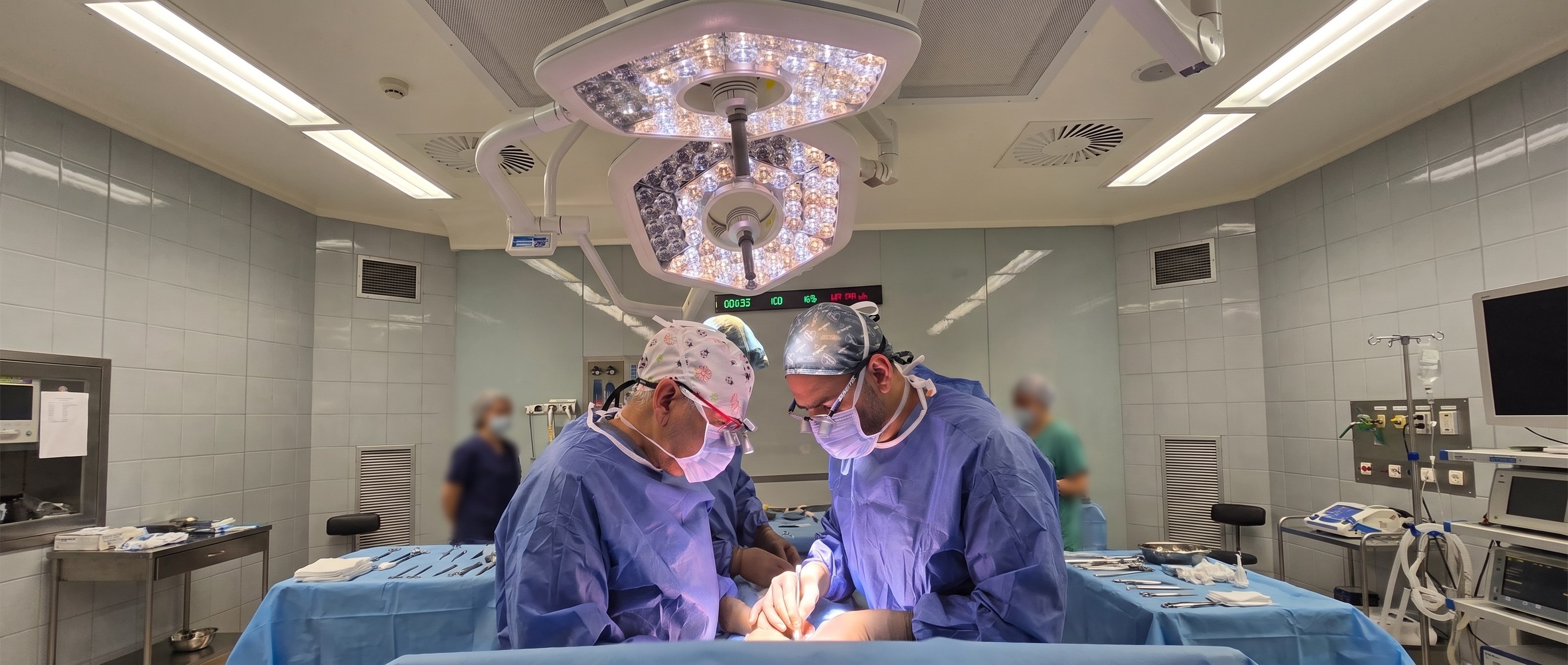
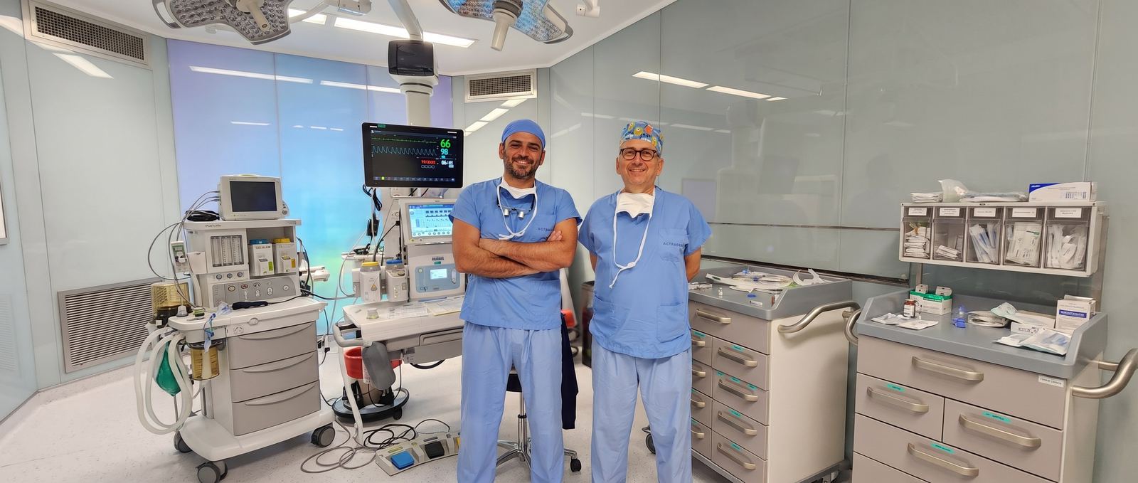
Two pediatric urology specialists work together on every operation: one performs the repair while the other assists, checks, and takes over when needed. This built-in second opinion completes demanding operations with less risk.

  • Lower risk

    A shared assessment and four-handed technique; a second experienced pair of eyes at the demanding steps.

  • Shorter surgery

    Working in parallel shortens the time spent under anesthesia.

  • Less anesthesia

    Infants and young children spend less time under anesthesia.

Prof. Dr. Ali Avanoğlu — hypospadias surgeon, Turkey

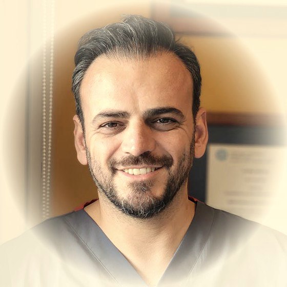
Prof. Dr. Ali Avanoğlu

Pediatric Surgeon and Pediatric Urologist

Over forty years in medicine, thirty-two of them as a pediatric surgeon; focused exclusively on pediatric urology since 1994. Associate professor in 1996 and full professor in 2002 at Ege University, where he chaired both the Department of Pediatric Surgery and the Division of Pediatric Urology. Roughly 3,000 hypospadias repairs and more than 1,000 operations for vesicoureteral reflux.

Doç. Dr. Yaşar Issı — hypospadias surgeon, Turkey

Doç. Dr. Yaşar Issı

Pediatric and Adult Urologist

Graduate of Ege University Faculty of Medicine (2006); completed urology training at İzmir Atatürk Training and Research Hospital (2011) and a pediatric urology fellowship at Ondokuz May University (2016); associate professor since 2022. Has worked at Gaziantep Children's Hospital, İzmir Çiğli Training and Research Hospital, and Bakırçay University. More than 1,000 hypospadias repairs and a comparable number of stone operations; continues the same patients' follow-up from childhood through adolescence and adulthood, in the same team.

Why together?

What operating together means for your child

In every operation, it is not a surgeon and an assisting resident — it is two specialists who both know the procedure, working together. We summarize the difference in four points.

Difference 01

Critical decisions are made together

Which technique to use, whether the repair will finish in one session, and how to respond to anything unexpected during surgery are decided by two specialists' shared assessment — never by one person alone.

Shared decision
Two pediatric urology surgeons
Together, in the same team, every time
Two hypospadias surgeons operating together
Difference 02

Shorter time under anesthesia

While one surgeon finishes a step, the other prepares the next; four hands work at once. In an infant or young child, every extra minute under anesthesia is a risk in its own right.

Two hands, at the same time
Difference 03 Related conditions, same session
1×

One anesthesia: conditions found together (undescended testis, inguinal hernia, hydrocele, hydronephrosis) are planned into the same session.

Get in touch
Difference 04 — Follow-up never breaks off

Some issues only surface during adolescence or adulthood. Because the team includes both pediatric and adult urology, follow-up never breaks off between childhood and adulthood.

Pediatric urology
Adult urology
Same team, same record
Process · for patients travelling from abroad too

Travelling to Turkey for treatment?

Surgery takes place at İzmir Acıbadem Kent Hospital; our clinic is about 4–5 km from the hospital, with a range of accommodation options nearby. The process runs in coordination between the clinic and the hospital, from your first consultation through to the completion of post-operative checks.

Clinic Hospital İzmir Acıbadem Kent Hospital · about 4–5 km.
  1. 01 Clinic
    Pre-treatment review with two pediatric urology surgeons — family on a video call from home

    Consultation and surgical planning

    Patients are first assessed at our clinic; surgical planning is done here.

    Avanoğlu–Issı Clinic

  2. 02 Hospital
    Prof. Dr. Ali Avanoğlu and Doç. Dr. Yaşar Issı, hypospadias surgeons in İzmir, Turkey

    Pre-operative tests and anesthesia review

    One day before surgery, you are referred to İzmir Acıbadem Kent Hospital for the required tests and anesthesia assessment.

    One day before surgery

  3. 03 Hospital
    Two pediatric urology surgeons' treatment process — family arriving at the hospital

    Surgery

    The operation takes place the next day, as planned. In suitable cases, it is completed as day surgery.

    İzmir Acıbadem Kent Hospital

  4. 04 Clinic
    Two pediatric urology surgeons' treatment process — remote follow-up after surgery, mother on a video call from home

    Recovery after discharge

    After the observation period, patients who meet discharge criteria continue recovering at home or at their accommodation. Dressing changes, wound checks, and catheter checks and removal continue at our clinic.

    Avanoğlu–Issı Clinic

Frequently asked by families

What families travelling from abroad ask first

Short answers are below; write to us directly with any question about your trip.

Let's plan your trip together
  • İzmir, Turkey
  • Two pediatric urology specialists
  • Same team from diagnosis to follow-up
Where do the operations take place?
Surgery takes place at İzmir Acıbadem Kent Hospital. Our clinic is about 4–5 km from the hospital, with a range of accommodation options nearby.
How does the process work, from consultation to surgery?
You are first assessed at our clinic, where the surgical plan is made. One day before surgery you are referred to the hospital for the required tests and anesthesia review; the operation itself takes place the following day, as planned.
Do we stay in hospital after surgery?
A significant share of our operations, when suitable, are completed as day surgery. Patients who meet discharge criteria continue recovering, after the observation period, in the more comfortable setting of your home or accommodation.
How are post-discharge checks handled?
Follow-up steps such as dressing changes, wound checks, and catheter checks and removal continue to take place at our clinic. That way, the entire process — from the first consultation through to the completion of follow-up — runs in coordination between the clinic and the hospital.
Topic index • 7 clusters

All UPJ obstruction topics

7 clusters, 45 detail pages. Each page focuses on a single question and links back here, to the main treatment page.

After surgery and follow-up6

Care, JJ stent, check-ups, possible problems, recurrent narrowing, and endoscopic methods.

Scientific publications · 4 articles

Prof. Dr. Ali Avanoğlu and Doç. Dr. Yaşar Issı's publications on UPJ Obstruction

Articles indexed in PubMed; the complete set of citations from the pages above. For all publications, see the Publications page →

  1. Avanoglu A, Tiryaki S (2020). Embryology and Morphological (Mal)Development of UPJ. Frontiers in pediatrics. PubMed ↗
  2. Kantar M, Hekimgil M, Harman M, et al. (2019). Unusual presentation of diffuse large B-cell non-Hodgkin's lymphoma in children: bilateral ureteral involvement. Pediatric hematology and oncology. PubMed ↗
  3. Resorlu B, Issi Y, Onem K, et al. (2016). Management of lower pole renal stones: the devil is in the details. Annals of translational medicine. PubMed ↗
  4. Tiryaki S, Alkac AY, Serdaroglu E, et al. (2013). Involution of multicystic dysplastic kidney: is it predictable? Journal of pediatric urology. PubMed ↗
Contact us

With the UPJ obstruction assessment that fits your situation would you like to reach the doctor?

There are two ways. If you'd like the doctor to already know your situation before replying, start with the short assessment; if your question is brief, write to us directly. Both reach the same team.

1 Let me assess my situation firstRecommended The Roadmap asks a few questions in about 2 minutes, collects your answers with an anatomical diagram, and produces a ready-made summary. That summary is added to your WhatsApp message — the doctor replies already knowing your situation. Start the Roadmap
2 I'll write directly Fill in the form; your message opens ready-made in WhatsApp (you can edit it before sending), or you can send it directly by email. This page's topic is added to the message automatically.

Editor's note: Age and a short sentence are enough; you don't need to use medical terms. Please don't send photos — the doctor will request them through a secure channel if needed. What you write is only sent when you submit it; this page does not store anything.

Direct contactReply comes from the doctor
Who is this for?
Quick questions:

Your message is not stored on our server; it is sent by WhatsApp/email. Privacy notice →

Gomauna Web Design and Development gomauna.com Prof. Dr. Ali Avanoğlu | 2026 © All Rights Reserved. SEO & GEO OPTIMIZED
WhatsApp Roadmap