Skip to content
Prof. Dr. Ali Avanoğlu
Main treatment page • Vesicoureteral Reflux (VUR)

Vesicoureteral Reflux (VUR) Treatment in Turkey

Vesicoureteral reflux (VUR) is the backward flow of urine from the bladder up through the ureter toward the kidney, during voiding or as the bladder fills. In children, it is most often discovered after a febrile urinary tract infection, or while investigating kidney enlargement found before birth. Treatment isn't a single method — it's a set of pathways that vary with grade, age, infection history and the condition of the kidney; spontaneous resolution is expected at low grades, and antibiotic prophylaxis, endoscopic injection or surgery come into play when needed. This page walks through that whole picture.

ByProf. Dr. Ali AvanoğluUpdated
At a glance 43 pages • 14 video
~30–40%
VUR in febrile UTI

Guideline range; found in a share of children with a febrile infection

10–80% depending on grade
Spontaneous resolution

Over the years, with growth; highest in low grades

Day surgery
Injection

~70–80% resolution after first session; performed jointly by two specialists

The whole treatment • 12 sections

This page explains the whole picture; the details of each topic are in the pages below

Start with what vesicoureteral reflux is and why it happens; then symptoms, the path to diagnosis, grades, its effect on the kidney, the bladder-bowel connection, the treatment decision, surveillance and antibiotic prophylaxis, endoscopic injection, reimplantation, and follow-up: twelve sections. Each section has detail pages below it; jump to the one you want from the contents on the right (at the top on mobile).

vesicoureteral reflux
01Section 1 / 12

Vesicoureteral reflux (VUR): what is it?

Urine flows backward from the bladder toward the kidney; most children are found through a febrile infection

Vesicoureteral reflux (VUR), commonly called kidney reflux, is a disruption of the urinary tract's normally one-way flow. The kidneys produce urine, which travels down through thin channels called ureters into the bladder, where it collects; as the bladder fills and empties, a natural valve mechanism where the ureters enter the bladder wall normally prevents urine from flowing backward. When this valve does not work well enough, urine flows backward from the bladder into the ureter and toward the kidney. In everyday language this is sometimes called "urine backflow" or "urine leaking up to the kidney."

RIGHTLEFT
Grade III reflux on the left: the red flow is urine flowing back from the bladder to the kidney; on the right, normal downward flow
Definitionin three sentences
  • 1Urine flows backwardas the bladder fills or empties, urine rises up into the ureter and toward the kidney
  • 2It gives no symptom of its ownit's mostly noticed through a febrile urinary tract infection or kidney enlargement seen before birth
  • 3The real concern is the kidneythe goal is to keep infection from reaching the kidney and leaving a scar
Quick factsat a glance
Frequency~1% of all children; ~25–40% of those with a febrile UTI
DiagnosisVCUG; ultrasound alone cannot give a grade
CourseMost grade I–II cases resolve on their own
TreatmentSurveillance, antibiotic prophylaxis, endoscopic injection, reimplantation
TeamDecisions and procedures with two specialists; follow-up continues into adulthood
Not to be confused with urinary incontinence

Vesicoureteral reflux is not urine leaking out of the body — it's urine flowing backward from the bladder toward the kidney; bedwetting is a separate topic.

Read the full text
  • VUR occurs in roughly 1% of the general child population; among children who have had a febrile urinary tract infection (UTI), the reported rate in the literature is 25–40%. Among babies found to have kidney enlargement before birth (antenatal hydronephrosis), around 10–20% also have reflux. Girls are more often diagnosed after an infection, while boys stand out in infancy and among higher grades.
  • Reflux itself doesn't cause pain and doesn't directly bother the child. What makes it important is that it makes it easier for bacteria in the bladder urine to be carried up to the kidney, setting the stage for a febrile kidney infection (pyelonephritis). Repeated kidney infections can leave some children with permanent scarring in the kidney tissue; the whole purpose of treatment is to break this chain.
  • Two concepts that families often confuse should be separated from the start. Here, "backflow" is an internal, invisible leak of urine from the bladder toward the kidney; a child wetting themselves — urinary incontinence (enuresis) — is a completely different matter. Likewise, "reflux" often brings acid reflux to mind, but it has nothing to do with vesicoureteral reflux; that's why we consistently use the full term "vesicoureteral reflux" throughout this site.
  • The good news is that in most children, VUR resolves on its own over time, as the child grows. The treatment decision is less about "does reflux exist or not" and more about "what is the least invasive path that will protect this particular child's kidneys." Every section on this page is part of answering that question.
Why does it matter?

Although reflux itself may look harmless, it lets bacteria in the bladder reach the kidney and sets the stage for febrile kidney infection. Repeated infections can leave permanent scarring in the kidney tissue in some children; treatment and follow-up exist to keep that possibility to a minimum.


Videos in this sectionFull guide (14) →
Why does VUR happen? Primary and secondary VUR
02Section 2 / 12

Why does VUR happen? Primary and secondary VUR

The problem is usually the ureter's short tunnel in the bladder wall

The point where the ureter enters the bladder is called the ureterovesical junction (UVJ). The ureter passes through an oblique tunnel within the bladder wall before opening into the bladder cavity; as the bladder fills and stretches, this tunnel flattens and compresses the ureter shut. This is a purely anatomical one-way-flow mechanism that needs no muscle or valve structure of its own. Whether the tunnel is long enough — normally about five times the ureter's diameter — is what determines whether the valve works.

URETERINSIDE THE BLADDERLONG TUNNELNORMAL: long tunnel, closes when the bladder fillsURETERINSIDE THE BLADDERSHORT TUNNELVUR: short tunnel, urine flows back to the kidney
A normal long tunnel closes when the bladder fills; in a short tunnel, urine flows back to the kidney
Two typesprimary / secondary
Primary VURthe valve itself falls short
  • The ureter's tunnel in the bladder wall is short
  • Congenital; familial tendency
  • The tunnel lengthens with growth → resolution
Secondary VURbladder pressure is high
  • Posterior urethral valves, neurogenic bladder
  • Voiding dysfunction, constipation (BBD)
  • The underlying problem is treated first
How the valve worksthree steps
  1. 1The ureter enters the wall at an anglepassing through a tunnel inside the bladder muscle
  2. 2As the bladder fills, the tunnel is compressedit closes like a valve; urine cannot rise back up
  3. 3If the tunnel is too short, it doesn't closeurine flows backward — reflux
Read the full text
  • The most common cause of VUR is that this tunnel is congenitally short, or that the ureteral opening sits further to the side and higher than it should. This is called primary VUR, and it's the type seen in the large majority of children. It is present from birth, has nothing to do with anything the family did or didn't do, and a significant share resolves on its own as the child grows, because the bladder wall thickens and the tunnel lengthens.
  • Secondary VUR describes situations where the valve mechanism is structurally normal but is overwhelmed by abnormally high pressure inside the bladder. This group includes posterior urethral valves (PUV), which narrow the urinary channel in male infants, neurogenic bladder due to spinal cord problems, and severe voiding dysfunction that sets in around toilet-training age. In secondary reflux, treatment first targets the bladder and the underlying cause; once bladder pressure drops, the reflux often recedes on its own.
  • In some children, reflux is accompanied by anatomical differences: a duplex collecting system, where two separate ureters arise from the same kidney (sometimes called a "double kidney" in everyday language); a ureterocele, where the ureteral opening balloons into the bladder; or a paraureteral diverticulum, a pouch that forms in the bladder wall next to the ureteral opening. In these situations, the chance of spontaneous resolution is lower and planning proceeds differently.
  • VUR has a familial pattern. Reflux has been reported in around 25–30% of siblings of a child with reflux, and in around 30–35% of children of a parent with a history of reflux; most of these are low-grade and silent. This information isn't meant to worry families — it matters because it helps the physician think of reflux earlier if a sibling develops a febrile infection.
Did we do something wrong?

No. Primary VUR is a congenital anatomical difference; it is not the result of anything done or not done during pregnancy, of diet, or of infant care. There can be a familial tendency, but that isn't anyone's responsibility either.

Videos in this sectionFull guide (14) →
Prof. Dr. Ali Avanoğlu's publications on this topic · 1
  1. Celik A, Ulman I, Aydin M, et al. (2002). Familial vesicoureteral reflux in asymptomatic siblings. The Turkish journal of pediatrics. PubMed ↗
UTI symptoms in babies
03Section 3 / 12

Symptoms of VUR: how is it noticed?

Reflux itself gives no symptom; a febrile urinary tract infection is what gives it away

VUR has no symptom of its own; the child doesn't feel it and it causes no pain. That's why reflux is almost always uncovered through some other event. The most common route is a febrile urinary tract infection: when an infection that starts with unexplained high fever, fussiness, vomiting, poor feeding and failure to gain weight in an infant or young child turns out to have bacterial growth on urine culture, the physician investigates the possibility of reflux.

How is it noticed?four routes
  • 1Febrile urinary tract infectionthe most common; unexplained fever, vomiting, fussiness in an infant
  • 2Kidney enlargement seen before birthpostnatal ultrasound and, if needed, VCUG
  • 3Family historya sibling or parent with reflux → ultrasound first
  • 4Silent refluxmay give no symptom at all; found incidentally on ultrasound
Fever in an infant

For fever of unknown cause, a urinalysis and a properly collected culture are the first step; a febrile infection can reach the kidney.

Read the full text
  • In older children, a urinary tract infection produces more familiar symptoms: burning on urination, frequent small-volume urination, foul-smelling or cloudy urine, lower abdominal pain, and new-onset daytime or nighttime wetting. When fever and flank pain are added to these symptoms, the infection may have reached the kidney (pyelonephritis); this picture is more serious and requires prompt treatment.
  • The second route is prenatal ultrasound. In some babies found during pregnancy monitoring to have kidney enlargement (antenatal hydronephrosis), the work-up done after birth finds VUR. These babies have never had an infection yet; the goal for them is to prevent the first infection and protect kidney development.
  • The third route is family history: in a child whose sibling or parent is known to have had reflux, the physician may investigate with a lower threshold. Guidelines recommend starting with ultrasound in these children; a voiding cystourethrogram (VCUG) is only considered for siblings with an abnormal ultrasound or a history of infection. In some children, reflux is found entirely by chance, on imaging done for another reason.
  • The practical point families should know is this: in a child under two, any fever without an obvious cause deserves evaluation for a urinary tract infection. Urinalysis and a properly collected urine culture are the starting point for both diagnosing the infection and investigating reflux; it's important to know that bag-collected samples can be misleading.
Take febrile UTI seriously

In infants and young children, unexplained high fever, vomiting, fussiness and poor feeding; in older children, flank pain, chills and cloudy urine alongside fever, can be signs of a kidney infection. Don't wait it out — it's important that antibiotics not be started before a urine culture is taken, so that later evaluation can be done properly.

Videos in this sectionFull guide (14) →
urinary tract infection in children
04Section 4 / 12

How is VUR diagnosed? Urine culture, ultrasound, VCUG and DMSA

Ultrasound sees it, VCUG confirms it, DMSA assesses the kidney

Diagnosing VUR proceeds in steps, and not every child gets the same full set of tests. The first step is the infection itself: urinalysis and a urine culture collected the right way (catheter or clean-catch midstream in an infant, never a bag sample) confirm a febrile UTI. Everyone then gets a kidney-bladder ultrasound; ultrasound shows kidney size, whether there's any dilation, the bladder wall, and post-void residual urine. The important point to know, however, is that a normal ultrasound does not rule out reflux, and ultrasound cannot tell you the grade of reflux.

RIGHTLEFT
VCUG: contrast is given into the bladder via a catheter; if reflux is present, it rises into the ureter and kidney
Testswho, what, when
TestWhat it showsWhen
Urinalysis + cultureWhether there's an infection; which organismFirst step in a febrile child; a proper sample is essential
Kidney-bladder ultrasoundDilation, kidney size, bladder wallIn every child with a febrile UTI
VCUGWhether reflux is present, its grade, which side; the urethraUnder 2 years, recurrent/atypical UTI, abnormal ultrasound, family history
DMSA scanKidney scarring and relative functionSelected cases: suspected scarring; 4–6 months after a febrile UTI
Two routesbottom-up / top-down
  1. 1Bottom-upfebrile UTI → ultrasound → VCUG (the classic route)
  2. 2Top-downfebrile UTI → acute DMSA; VCUG can be skipped if there's no kidney involvement (center-dependent)
  3. 3Radiation-free alternativecontrast-enhanced voiding urosonography (ceVUS), especially in girls and for follow-up
Read the full text
  • The test that definitively shows reflux is the voiding cystourethrogram (VCUG). Contrast is instilled into the bladder through a thin catheter, and X-ray images are taken as the bladder fills and as the child voids, showing whether the contrast backs up into the ureter and kidney, and if so, how far up it goes. Grade is determined only by this test. The procedure takes 20–30 minutes, the radiation dose is low, and it is done after the infection has cleared, if needed under single-dose antibiotic cover.
  • VCUG is not done for everyone. Guidelines recommend it after a febrile infection particularly for: children under two, those with recurrent, atypical or severe infections, those with an abnormal ultrasound, those with a family history of reflux, and male infants. The American Academy of Pediatrics, by contrast, does not routinely recommend VCUG after a first febrile infection if the ultrasound is normal, reserving it for a second febrile infection. Between these two approaches, the physician decides based on the child's age, the severity of the infection, and the ultrasound findings.
  • Some centers use a "top-down" approach: after a febrile infection, a DMSA renal scan first checks whether the kidney is affected, and VCUG can be skipped if there's no kidney involvement. This approach reduces the number of VCUGs but can miss low-grade reflux; which path is followed depends on the center and the case. As a radiation-free option, contrast-enhanced voiding urosonography (ceVUS) is also being used increasingly, particularly in girls and for follow-up; it is available at a limited number of centers.
  • DMSA (a renal scan) does not show reflux — it shows the condition of the kidney tissue itself: how well each kidney is functioning and whether there is scarring. Reduced uptake seen right after a febrile infection can be temporary; scanning is done at least 4–6 months after the infection to assess for permanent scarring. Ultrasound, VCUG and DMSA do not substitute for one another — each answers a different question, and which child needs which test is decided by the same team, from diagnosis through treatment.
Does VCUG harm the child?

VCUG is a short, low-radiation-dose test that children tolerate well in experienced hands; there is brief discomfort while the catheter is placed. It is done after the infection has cleared, with single-dose antibiotic cover given when needed. Preparing families in advance, and explaining the test to the child in an age-appropriate way, makes the procedure noticeably easier.

Videos in this sectionFull guide (14) →
VUR grades: what do grades I to V mean?
05Section 5 / 12

VUR grades: what do grades I to V mean?

Five grades: from the ureter alone to marked dilation

VUR is classified into five grades, I through V, using the International Reflux Study Committee system. Grade is determined by how far up the contrast rises on VCUG and how much it dilates the ureter and the kidney's collecting system. This classification is more than a label — it directly affects the likelihood of spontaneous resolution, the risk of infection and kidney damage, and the treatment options.

RIGHTLEFT
Grade I · ureter only
RIGHTLEFT
Grade II · reaches the pelvis, no dilation
RIGHTLEFT
Grade III · mild dilation
RIGHTLEFT
Grade IV · marked dilation, tortuous
RIGHTLEFT
Grade V · severe dilation, blunted calyces
Five grades side by side: I ureter only → V severe dilation and tortuous ureter
Five gradesby VCUG
GradeAppearanceSpontaneous resolution (approx.)
IUreter only; no dilation70–80%
IIReaches the renal pelvis; no dilation70–80%
IIIMild-to-moderate dilation; calyces mildly blunted~50%
IVMarked dilation; ureter tortuous~30%
VSevere dilation; calyces blunted, ureter very tortuous10–15%
Read the full text
  • In grade I, urine backs up only into the ureter and does not reach the kidney, with no dilation. In grade II, urine rises all the way to the kidney's collecting system, but neither the ureter nor the renal pelvis is dilated. These two grades are called "low-grade" or "non-dilating" reflux; they have the highest rate of spontaneous resolution and are mostly followed with observation alone.
  • In grade III, there is mild-to-moderate dilation of the ureter and renal pelvis, with mild blunting of the calyces (the small collecting cups inside the kidney). In grade IV, dilation becomes more pronounced and the ureter starts to become tortuous. In grade V, the ureter is markedly dilated and tortuous, and the renal pelvis and calyces are severely dilated. Grades III–V are referred to as "dilating" reflux, and treatment decisions are made more actively in this group.
  • The relationship between grade and spontaneous resolution is clear: the literature reports cumulative resolution over the years at roughly 70–80% for grades I–II, around 50% for grade III, around 30% for grade IV, and 10–15% for grade V. These figures are averages; bilateral disease, diagnosis at an older age, bladder-bowel dysfunction, recurrent infection and existing scarring all lower the chance of resolution.
  • The question families ask most is "will the grade get worse?" In primary reflux, grade does not increase over time — on the contrary, it decreases or disappears with growth. The same child can have different grades on the two sides, and one-sided reflux tends to have a better course than two-sided reflux. Grade alone doesn't dictate the decision; together with age, sex, infection history, kidney status and bladder function, it builds a "risk profile."
Ultrasound cannot tell you the grade

Seeing "mild kidney dilation" on ultrasound does not translate into a reflux grade — grade is determined only by VCUG. Even children with a completely normal ultrasound can turn out to have grade III–IV reflux; that's why the decision is based on the whole picture, not on a single test.

Videos in this sectionFull guide (14) →
Kidney damage: reflux nephropathy and renal scarring
06Section 6 / 12

Kidney damage: reflux nephropathy and renal scarring

What's really being protected is the kidney tissue: scarring, blood pressure and function

The real significance of VUR lies in its possible effect on the kidney. Reflux makes it easier for bacteria in the bladder to be carried up to the kidney; during a febrile kidney infection (pyelonephritis), inflammation develops in the kidney tissue, and some areas can heal with permanent scarring. This permanent mark in the kidney tissue is called a scar, and the overall picture of reflux-related kidney damage is called reflux nephropathy. On a DMSA scan, scarring shows up as an area of the kidney with no uptake.

RIGHT%62RELATIVE FUNCTIONLEFT%38RELATIVE FUNCTIONDashed rings: multiple scarred areas
DMSA scintigraphy: scarred areas in the left kidney and reduced relative function
Two kinds of scarringthe distinction matters
  • 1Congenital dysplasiamale, high grade, prenatal diagnosis; present even without infection
  • 2Acquired scarringrecurrent febrile infection; more common in girls; preventable
Watched over the long termwhy follow-up matters
Blood pressurerisk rises in adulthood with bilateral scarring
Proteinuriaannual urinalysis
Kidney growthby ultrasound
FunctionDMSA / blood tests when needed
Kidney failure?

End-stage kidney failure from reflux nephropathy has become rare and is mainly seen with severe, bilateral scarring; early diagnosis and preventing infection are the real protection.

Read the full text
  • Not every scar comes from infection — this distinction matters for families. In one group of children — particularly male infants with high-grade reflux found to have hydronephrosis before birth — the kidney formed incompletely during development itself (congenital dysplasia), and the DMSA findings are present even without ever having had an infection. In the second group, scarring is acquired later, after repeated pyelonephritis episodes; this picture is seen more in girls and after toilet training. In the first group, treatment aims to protect the tissue that remains; in the second, it aims to prevent new infections.
  • Several factors raise the risk of scarring: high grade, febrile infection at a young age and infections that recur, late start of treatment, bladder-bowel dysfunction, and bilateral disease. Conversely, recognizing a febrile infection early and treating it promptly markedly lowers the chance of scarring. This is why fever in a child known to have VUR is not a "wait and see" situation — a urine culture should be taken and treatment started without delay.
  • The long-term consequences of scarring depend on its size and on whether it is one-sided or two-sided. A small, one-sided scar usually causes no problems at all; extensive, two-sided scarring can, in later years, lead to high blood pressure, protein leaking into the urine, and rarely a decline in kidney function. The risk of hypertension in adulthood has been reported at around 10–20% in cases with bilateral scarring. End-stage kidney failure due to reflux nephropathy has become increasingly rare and is mainly seen in severe, two-sided scarring.
  • This information isn't meant to frighten — it's meant to explain why follow-up is spread over years. In children with scarring, annual blood pressure measurement, urinalysis and ultrasound, plus a DMSA scan within 4–6 months of a febrile infection, are the main parts of follow-up. That settles the question of whether VUR is "dangerous": it isn't the reflux itself that's dangerous — it's infections that go unprevented and scarring that goes unnoticed.
Does scarring reverse?

Once scarring has set into the kidney tissue, it does not reverse — but the formation of new scarring can largely be prevented. Early treatment of febrile infection, antibiotic prophylaxis in the right child, correcting bladder-bowel function, and correcting reflux when needed all serve this goal.

Videos in this sectionFull guide (14) →
Prof. Dr. Ali Avanoğlu's publications on this topic · 1
  1. Ozel SK, Dokumcu Z, Akyildiz C, et al. (2007). Factors affecting renal scar development in children with spina bifida. Urologia internationalis. PubMed ↗
Constipation, voiding dysfunction and reflux: bladder-bowel dysfunction (BBD)
07Section 7 / 12

Constipation, voiding dysfunction and reflux: bladder-bowel dysfunction (BBD)

Reflux treatment stays incomplete until constipation and voiding dysfunction are corrected

In children who have completed toilet training, one of the factors that most affects the course of VUR is how the bladder and bowel are working. Constipation, holding urine, waiting until the last moment when the urge strikes, being unable to relax the pelvic floor muscles while voiding, and not fully emptying the bladder are together called "bladder-bowel dysfunction" (BBD). This picture is frequently found alongside reflux, and the two feed each other.

CONSTIPATION: a full bowel presses on the bladderBLADDERBOWEL (RECTUM)REGULAR EMPTYING: the bladder fills and empties easilyBLADDERBOWEL (RECTUM)
With constipation a full bowel presses on the bladder; with regular emptying the bladder fills and empties easily
Does any of this apply?after toilet training
  • 1Urgency / incontinencean overactive bladder
  • 2Holding urine, leg-crossingholding maneuvers
  • 3Voiding too often or too rarelyfewer than 3 times a day / more than 8
  • 4Weak, interrupted streamdysfunctional voiding
  • 5Constipation, soilinga full rectum presses on the bladder
Priority: bladder and bowelurotherapy
  1. 1Timed voidingevery 2–3 hours, unhurried, feet supported
  2. 2Fluid routinewater spread through the day; less in the evening
  3. 3Treating constipationfiber, fluids, PEG if needed; a regular toilet time
  4. 4Further evaluation if neededvoiding diary, uroflowmetry, residual urine, biofeedback
Read the full text
  • The mechanism is straightforward: a full bowel presses on the bladder from outside, a tightened pelvic floor prevents the bladder from emptying fully, and the urine left behind becomes a breeding ground for bacteria. In a bladder that voids under high pressure and without emptying completely, reflux is easier to maintain and infection recurs. A combined analysis of the RIVUR and CUTIE trials found infection recurrence to be highest (in roughly half) in children with both reflux and BBD together, and lowest in children with neither.
  • That's why the shared message across guidelines is this: in every child who has completed toilet training, BBD is screened for — and treated if present — before deciding on reflux treatment. Screening uses simple tools: a detailed history and questionnaire with the family, a voiding diary kept over a few days, uroflowmetry, and ultrasound measurement of post-void residual urine. When BBD is present, the success of endoscopic injection or surgery drops and infections keep recurring; once BBD is corrected, the chance of reflux resolving on its own goes up.
  • The first step in treatment is urotherapy: going to the toilet at regular intervals (roughly every two to three hours), voiding unhurried and sitting with the feet supported, adequate fluid intake, and correcting constipation through diet and, if needed, medication. In most children, these simple changes make a noticeable difference within a few months. When that isn't enough, biofeedback, which teaches the child to relax the pelvic floor muscles, is added, along with bladder medication in selected children; antibiotic prophylaxis may accompany this phase in a child who also has reflux.
  • The practical message for families: if your child has been diagnosed with reflux and has completed toilet training, constipation and toilet habits matter at least as much as the grade of reflux. Share signs like leg-crossing, squatting, holding on while fidgeting, dampness in the underwear, or hard stools every few days with your physician — these can change the treatment plan.
Bladder and bowel first, then reflux

In a child who has completed toilet training, constipation and voiding habits are corrected before injection or surgery for reflux is even considered. When this order is followed patiently, some children need no procedure at all; and when a procedure is needed, its success rate rises markedly.

Videos in this sectionFull guide (14) →
Prof. Dr. Ali Avanoğlu's publications on this topic · 3
  1. Tiryaki S, Eraslan C, Soyer T, et al. (2019). Nonneuropathic Neuropathic Bladder-Is it Really Nonneuropathic? The Journal of urology. PubMed ↗
  2. Ural Z, Ulman I, Avanoglu A (2008). Bladder dynamics and vesicoureteral reflux: factors associated with idiopathic lower urinary tract dysfunction in children. The Journal of urology. PubMed ↗
  3. Sözübir S, Celik A, Emir N, et al. (2005). Consistency of urodynamic parameters in children with detrusor instability: how many times should the bladder be filled? Urologia internationalis. PubMed ↗
vesicoureteral reflux treatment
08Section 8 / 12

Which treatment for whom? Risk profile and the decision

The decision rests on eight factors read together, not a single number

There is no single correct treatment for VUR — the right one is whichever path fits that particular child's risk profile. Guidelines build this profile from the answers to a few questions: how old is the child and what sex? What is the grade and side of the reflux? Has the child had a febrile infection, how many times, and was it while on antibiotic prophylaxis? Is there scarring or loss of function in the kidney? If toilet training is complete, how is bladder-bowel function? These answers place the child roughly into a low, moderate or high risk group.

VUR gradeFebrile UTIBilateralKidney scarringLoss of functionBBD / voidingAgeAdditional anatomical issue
Eight risk factors; the red boxes are the ones that most influence the decision
Risk groupsthe EAU approach
GroupWhoFirst step
LowToilet-trained, grade I–III, one-sided, no/single UTI, no scarringSurveillance; culture with fever; treat BBD first if present
ModerateGrade III–IV over age 1, or all grades in a young infant, with a UTI historyAntibiotic prophylaxis ± urotherapy; injection if no improvement
HighGrade IV–V, bilateral, scarring/loss of function, febrile UTI while on CAPAntibiotic prophylaxis, close to corrective treatment: injection or reimplantation
The decision is made with two specialists

Age, grade, side, infection history, scarring, voiding habits and the family's preference are all read together; no single finding on its own requires surgery.

Read the full text
  • The low-risk group generally includes children over one year old with low-grade (I–II), one-sided reflux, no scarring, and no infection or only a single infection. In this group, the first option is active surveillance without antibiotic prophylaxis: urine culture with fever, annual ultrasound, and checking for BBD. Children with high-grade, two-sided, scarred or recurrent-infection reflux make up the high-risk group; here, close follow-up is combined with antibiotic prophylaxis, and corrective treatment (injection or surgery) is discussed earlier. In the broad middle group in between, the decision is individualized based on age and course.
  • Age is one of the strongest drivers of the decision. Antibiotic prophylaxis is recommended for every infant under one year with reflux who has had a febrile infection; it may also be recommended, based on the PREDICT study, for infants under one with high-grade (III–V) reflux who have not yet had an infection. Circumcision in a male infant is discussed with the family as an additional option that lowers infection risk. Over the age of one, the course is determined more by infection recurrence and BBD; in a child past toilet training, bladder-bowel function is corrected first.
  • Certain situations move the decision directly toward a procedure: a febrile infection occurring while on antibiotic prophylaxis, new or progressive scarring on DMSA, high-grade reflux that fails to improve despite long follow-up, an accompanying anatomical anomaly (ureterocele, duplex system, diverticulum), and an informed family's preference. At this point the choice is mostly between same-day endoscopic injection and open or minimally invasive ureteral reimplantation; which one is right is decided together, based on grade, anatomy and the family's priorities.
  • In our practice, this whole decision is made and carried out by the same two pediatric urology specialists: from deciding which tests are needed for diagnosis, to starting antibiotic prophylaxis, to endoscopic injection, reimplantation, and years of follow-up — the same team knows the child throughout. In secondary reflux, the order is different: for posterior urethral valves or neurogenic bladder, bladder pressure and the underlying cause are treated first; reflux usually recedes as a result, and a direct anti-reflux operation is not the first step.
Grade alone doesn't make the decision

Two children with the same grade of reflux can be treated in different ways: in one, age, sex and infection history support observation; in the other, a single febrile infection while on antibiotic prophylaxis can bring a procedure forward. So there is no single answer to "what do you do for grade III" — the answer lies in the whole child.

Videos in this sectionFull guide (14) →
Does VUR resolve on its own? Active surveillance and antibiotic prophylaxis
09Section 9 / 12

Does VUR resolve on its own? Active surveillance and antibiotic prophylaxis

Most reflux resolves on its own with growth

Yes — VUR resolves on its own in a significant proportion of children. The reason is growth: as the child grows, the bladder wall thickens, the ureter's tunnel within the bladder wall lengthens, and the valve mechanism matures. Completing toilet training and normalizing bladder-bowel function also contribute to this process. Resolution happens over years, not months; it is faster and more common in low grades, slower and less common in high grades.

%0%25%50%75%100DIAGNOSIS1 YR2 YR3 YR4 YR5 YRSHARE OF CHILDREN WITH PERSISTING REFLUXGrade I–IIGrade IIIGrade IV–VApproximate literature trend; spontaneous resolution is more likely at lower grades and younger ages. Individual course varies.
Spontaneous resolution: high at grade I–II, moderate at grade III, low at grade IV–V
Active surveillancewhat it means
  • 1Annual ultrasoundkidney growth and dilation
  • 2Culture with feverurinalysis with any febrile illness
  • 3Toilet habitsBBD is screened for and treated
  • 4Routine repeat VCUG not requiredconsidered at 12–24 month intervals
Antibiotic prophylaxis (CAP)who needs it
<1 year + febrile UTIrecommended at every grade
<1 year, grade III–V, no UTIcan be recommended (PREDICT)
>1 year, low grade, no/single UTIsurveillance is an option
During BBD treatmenttemporary cover
Febrile UTI while on CAPstep up treatment
How much?

A quarter to a third of the treatment dose, a single dose at night; reviewed in 6–12 month blocks. RIVUR: cuts febrile infection recurrence roughly in half, with no shown difference in scarring.

Read the full text
  • Active surveillance is not simply waiting and doing nothing. In a child on surveillance, the family learns to recognize the signs of febrile infection and gets a urine culture promptly with any fever; the physician follows the kidney with annual ultrasound, blood pressure and urinalysis; and BBD is screened for and corrected once toilet training is complete. Routine repeat VCUG is not needed for low-grade reflux; a follow-up film is mostly considered only at 12–24 month intervals, and only if it would change the decision.
  • Antibiotic prophylaxis (CAP) aims to keep the urine bacteria-free by giving a single low dose each night — roughly a quarter to a third of the treatment dose. The most commonly used drugs are trimethoprim-sulfamethoxazole after two months of age, nitrofurantoin after infancy, and amoxicillin or a first-generation cephalosporin in newborns and young infants. The RIVUR trial showed that antibiotic prophylaxis cut the recurrence of febrile infection roughly in half (about one recurrence prevented for every eight children treated); on the other hand, it made no difference to the rate of new scarring and increased the rate of resistant bacteria. This two-sided result is the basis for the principle that antibiotic prophylaxis should go to "the right child," not to everyone.
  • Who gets antibiotic prophylaxis? The synthesis across guidelines is this: it is recommended for every infant under one year with reflux who has had a febrile infection; it can be recommended for infants under one with high-grade (III–V) reflux who have not had an infection; in children over one with low-grade reflux and no infection or a single infection, surveillance is a valid option; and it accompanies treatment of BBD in children who have it. The Swedish Reflux Trial showed that, in girls aged 1–2, antibiotic prophylaxis or endoscopic treatment was superior to surveillance alone, while there was no clear difference in boys — which is why sex also factors into the decision.
  • Duration is generally reviewed in 6–12 month blocks; it can be stopped once reflux resolves, toilet training is complete, or the child grows older. A febrile infection occurring while on antibiotic prophylaxis — a "breakthrough" infection — is an important warning sign: adherence to the medication and BBD are checked first, and then treatment is stepped up — most often to endoscopic injection or surgery.
Quick summary

Grades I–II: mostly surveillance, ~70–80% resolve over the years. Grade III: surveillance ± antibiotic prophylaxis, ~50% resolve. Grades IV–V: antibiotic prophylaxis and close follow-up; ~10–30% resolve, and a procedure comes up more often. Under one year + febrile infection: antibiotic prophylaxis. Febrile infection while on antibiotics: step up treatment.

Videos in this sectionFull guide (14) →
Prof. Dr. Ali Avanoğlu's publications on this topic · 1
  1. Ulman I, Avanoğlu A, Erikçi V, et al. (1998). Is resolution of vesicoureteric reflux by conservative management predictable in patients with myelodysplasia? European urology. PubMed ↗
Endoscopic injection treatment (Deflux, subureteral bulking)
10Section 10 / 12

Endoscopic injection treatment (Deflux, subureteral bulking)

Day surgery, no incision: a bulking agent placed below the ureteral opening

Endoscopic injection is the incisionless treatment for VUR. Under general anesthesia, a thin camera (cystoscope) is passed through the urethra into the bladder, and a special bulking agent (dextranomer/hyaluronic acid, brand name Deflux) is injected just below or into the refluxing ureteral opening. The resulting mound lifts the ureteral opening, reshapes it into a slit, and creates a tunnel-like effect that can close when the bladder fills. The procedure takes 15–30 minutes, is done as day surgery, and the child goes home the same day.

INSIDE THE BLADDER · CYSTOSCOPIC VIEWBEFORE: opening patent, refluxINSIDE THE BLADDER · CYSTOSCOPIC VIEWAFTER: bulking agent supports the opening, a valve forms
Endoscopic injection: before, an open opening with reflux; after, the bulking agent supports the opening
The procedurestep by step
  1. 1General anesthesia, cystoscopyno incision; a thin camera enters the bladder
  2. 2Bulking agent below the ureteral openingDx/HA (Deflux) with the STING/HIT technique; the opening reshapes into a slit
  3. 3Day surgery15–30 minutes; home the same day
  4. 4Follow-upultrasound at 4–6 weeks; VCUG/ceVUS at 3–6 months (center-dependent)
Successby grade, single session
GradeApproximateNote
I–II80–90%~85% with a repeat
III70–75%lower with BBD
IV60–65%a second session is an option
V30–50%reimplantation is more reliable
Read the full text
  • The injection technique — different needle placements known by names like STING and HIT — and delivering the right amount of bulking agent to the right spot are what determine success. In our practice, endoscopic injection is a procedure both pediatric urology specialists take part in together; the ureteral opening is assessed and the injection site is decided jointly. Because the same team also performs reimplantation when needed, a switch to the alternative, if injection turns out not to be suitable, is planned without any break in continuity.
  • Success varies by grade and shouldn't be described with a single number. In the literature, the rate of resolution after a single session is reported at roughly 80–90% for grades I–II, 70–75% for grade III, 60–65% for grade IV, and 30–50% for grade V; with a second injection, overall success reaches around 85%. Because late recurrence of around 10–20% has been reported in the long term, follow-up after treatment matters. Success drops in the presence of a duplex collecting system, posterior urethral valves, neurogenic bladder, and untreated BBD.
  • Who is a good candidate for injection? It is usually the first corrective option in children over one year old with low-to-moderate grade (II–IV) reflux who have had a febrile infection while on antibiotic prophylaxis, or who want to come off long-term antibiotics. In children with high-grade reflux, markedly dilated ureters, or anatomical anomalies, success rates drop and surgery becomes the more favored option; some families, meanwhile, prefer to try the less invasive option first. This choice is made together with the physician, discussed with honest numbers.
  • Side effects are rare: mild burning or a small amount of blood in the urine can be seen for a few days after injection; febrile infection is uncommon. Temporary or permanent ureteral obstruction has been reported at under 1% and is kept to a minimum by correct placement of the bulking agent. Follow-up involves an ultrasound at 4–6 weeks and, depending on the center, a VCUG or contrast-enhanced voiding urosonography at 3–6 months; if reflux persists, a second injection or surgery is discussed.
Is injection permanent?

The bulking agent stays in the body, and the effect is lasting in most children; however, the literature reports late recurrence at around 10–20%. That's why, if a febrile infection occurs after injection, reflux is investigated again — follow-up doesn't end with the injection.

Videos in this sectionFull guide (14) →
Prof. Dr. Ali Avanoğlu's publications on this topic · 3
  1. Tekin A, Yagmur I, Tiryaki S, et al. (2018). Changing bulking agent may require change in injection volume for endoscopic treatment of vesicoureteral reflux. International braz j urol. PubMed ↗
  2. Kocherov S, Ulman I, Nikolaev S, et al. (2014). Multicenter survey of endoscopic treatment of vesicoureteral reflux using polyacrylate-polyalcohol bulking copolymer (Vantris). Urology. PubMed ↗
  3. Ozcan C, Ergün R, Ozbek SS, et al. (2007). Bladder ultrasound in the evaluation of the efficacy of dextranomer/hyaluronic acid injection for treatment of vesicoureteral reflux. Journal of clinical ultrasound. PubMed ↗
life after vesicoureteral reflux surgery
11Section 11 / 12

VUR surgery: when is ureteral reimplantation needed?

Reimplantation: a new, longer tunnel for the ureter

The operation for VUR is called ureteral reimplantation: reconnecting the ureter to the bladder wall through a new tunnel that is long enough to work properly. The goal is to lengthen the congenitally short tunnel — to roughly five times the ureter's diameter — and rebuild the valve mechanism. This is the most definitive treatment for VUR; the literature reports a resolution rate of about 95–98% with open reimplantation.

COHEN · through the bladder, cross-trigonal tunnelNew opening on the opposite side; the tunnel lengthensLICH-GREGOIR · from outside the bladderThe ureter is laid into a groove and covered with muscle
Reimplantation: Cohen (through the bladder, cross-trigonal tunnel) and Lich-Gregoir (from outside the bladder)
Two approachesreimplantation
Intravesical (Cohen)cross-trigonal tunnel
  • The bladder is opened; a new, longer tunnel for the ureter
  • Success ~95–98%
  • 1–3 day stay; catheter
Extravesical (Lich-Gregoir)outside the bladder
  • The bladder isn't opened; the ureter is laid into a groove and covered with muscle
  • Can also be done laparoscopically/robotically (~88–97%)
  • Temporary voiding difficulty possible if done bilaterally
When does it come up?indications
  • 1Febrile UTI while on CAPprophylaxis isn't enough
  • 2New or progressive scarringthe kidney is being harmed
  • 3No improvement at a high gradeIV–V, over the years
  • 4Anatomical anomalyureterocele, duplex system, diverticulum
  • 5Injection has failedafter a second session
  • 6Family preferencewanting a definitive solution
Read the full text
  • Who needs surgery? The shared list across guidelines is: a febrile infection occurring while on antibiotic prophylaxis, new or progressive scarring or declining kidney function on DMSA, high-grade reflux that fails to resolve despite long follow-up, failed endoscopic injection, an accompanying anatomical anomaly (ureterocele, duplex collecting system, paraureteral diverticulum), and the preference of an informed family. Most of these items are weighed together; no single finding on its own triggers a decision for surgery.
  • There are two basic approaches. In the intravesical Cohen technique, the bladder is opened and the ureter is passed through a horizontal tunnel in the bladder wall toward the opposite side; it is often chosen for bilateral reflux. In the extravesical Lich-Gregoir technique, the bladder is not opened — the ureter is laid into a bed created on the outer surface of the bladder and covered with muscle tissue; catheter time and bladder spasm are generally less with this approach. Which technique is chosen depends on the side involved, the width of the ureter, and any accompanying anomaly.
  • The same principles can also be applied laparoscopically or with robotic assistance, through small openings in the abdominal wall. With these methods the incisions are smaller and the hospital stay is shorter, though operating time is longer, and success rates in the literature range from 88–97%, depending on center experience. Open surgery requires a 1–3 day hospital stay, a short-term bladder catheter, and, in selected cases, a ureteral stent; children mostly return to normal life within one to two weeks.
  • In our practice, ureteral reimplantation is always performed jointly by the two pediatric urology specialists; since the same two specialists also perform endoscopic injection, the choice of which path to take — and switching from one to the other if needed — is planned by the same hands throughout. Possible problems are uncommon: ureteral obstruction in roughly 1–2%, new reflux on the opposite side in roughly 5–10%, and temporary voiding difficulty with the bilateral extravesical technique. Most of these resolve when caught early, which is why post-operative checks are scheduled.
Is a follow-up VCUG needed after surgery?

After open reimplantation, a routine VCUG is not required if the follow-up ultrasound is normal and there has been no febrile infection; guidelines leave this as optional. After injection, a follow-up film or contrast-enhanced voiding urosonography at 3–6 months is more common. The decision is made according to the center and the individual case.

Videos in this sectionFull guide (14) →
Doç. Dr. Yaşar Issı's publications on this topic · 1
  1. Issi Y (2020). Non-Invasive Stent Removal after Ureteroneocystostomy in Pediatric Patients: Long-Term Results. Urology journal. PubMed ↗
After treatment and long-term follow-up: adolescence, adulthood, pregnancy
12Section 12 / 12

After treatment and long-term follow-up: adolescence, adulthood, pregnancy

Follow-up continues without a break from pediatric to adult urology

Whether VUR resolves on its own or is corrected with injection or surgery, it is not a file that closes with treatment. The backbone of follow-up is the same at every age: annual kidney-bladder ultrasound, blood pressure measurement and urinalysis (especially for protein leakage), and monitoring of growth. If a febrile infection occurs, it is confirmed by culture, treated promptly, and checked with a DMSA scan 4–6 months later for any new scarring. In unscarred, low-grade, resolved reflux, follow-up becomes less frequent over time and is ended once certain conditions are met.

FIRST 3 MONTHSUltrasound · urineFIRST YEARUS ± VCUG · blood pressureCHILDHOODAnnual checkADOLESCENT · ADULTPre-pregnancyPROCEDURE / DIAGNOSIS3 MO12 MOADOLESCENCEADULTOUTCOME ASSESSMENT (VCUG IF NEEDED)
Follow-up chart: first 3 months ultrasound · first year outcome assessment · annual check in childhood · adolescent and adult
Timelinefrom diagnosis to adulthood
First 3 monthsUltrasound and urine after the procedure; obstruction is ruled out after injection/surgery
First yearOutcome assessment: ultrasound, VCUG/ceVUS if needed; blood pressure
ChildhoodAnnual ultrasound, blood pressure, urinalysis; culture and DMSA (4–6 months) with febrile UTI
Adolescence · adulthoodBlood pressure in scarred patients; pre-pregnancy counseling for women; uninterrupted follow-up with the same team
Read the full text
  • After injection, follow-up is done with an ultrasound at 4–6 weeks and, depending on the center, a VCUG or contrast-enhanced voiding urosonography at 3–6 months; annual surveillance continues after that, and reflux is reassessed if a febrile infection occurs. After reimplantation, ultrasound is done at 4–6 weeks and again at about one year; a routine VCUG is not needed if the ultrasound is normal and there is no infection. After either treatment, toilet habits and constipation continue to be followed, because BBD keeps up infection risk independently of the treatment given.
  • Adolescence is a time for reassessment. If reflux is still present at this age, the chance of spontaneous resolution is low; but in a teenager with healthy kidneys, no scarring, and no infections, the risk of harm is also low, and observation alone is often sufficient. In adolescents with scarring, blood pressure and urine follow-up matter in particular; hypertension in this group can appear years later. The teenager should move into adulthood knowing their own situation.
  • Pregnancy is a separate topic for girls. Women with a history of VUR or scarring have a higher risk of urinary tract infection, kidney infection and elevated blood pressure during pregnancy; the risk relates more to the extent of scarring and kidney function than to the reflux itself. Routinely correcting persisting reflux before pregnancy is not supported by evidence; the decision is individual. Regular urine screening during pregnancy and early treatment of infection prevent most problems.
  • We place particular importance on follow-up running without interruption from childhood into adulthood: because Doç. Dr. Issı practices both pediatric and adult urology, post-adolescent checks, pre-pregnancy counseling, and questions that come up in adulthood are all answered from the same file. Having the childhood imaging, DMSA results and treatment history stay with the same team means that decisions made years later rest on solid ground.
When does follow-up end?

If reflux has resolved or been corrected, there is no kidney scarring, blood pressure and urine are normal, toilet habits are regular, and no febrile infections are occurring, follow-up is spaced out and usually concludes with an adolescent assessment. In those with scarring, annual blood pressure and urine checks continue into adulthood as well.

Videos in this sectionFull guide (14) →
Roadmap • Interactive tool

Assess your situation yourself

Answer a few questions; the anatomical diagram updates as you go. At the end you're guided to the section and pages that fit your situation, and you can send the summary to the doctor by WhatsApp. About 2 minutes; your answers stay on your device.

Vesicoureteral Reflux RoadmapStep-by-step assessment
Step 1
Loading tool…
Vesicoureteral Reflux (VUR) video guide • 14 video

What people ask about Vesicoureteral Reflux (VUR), in short videos

Prepared in order: first, what vesicoureteral reflux is, why it matters, and diagnosis; then treatment options and bladder-bowel habits; and finally follow-up after treatment.

Viewing order1–4 / 14
Frequently asked questions

The first 12 questions families ask

Short answers to the questions families ask most often.

Ask your question on WhatsApp
Does VUR resolve on its own?
In most children, yes. With growth, the ureter's tunnel within the bladder wall lengthens and the valve mechanism matures. The literature reports resolution over the years at roughly 70–80% for grades I–II, 50% for grade III, 30% for grade IV, and 10–15% for grade V. Bilateral disease, recurrent infection, bladder-bowel dysfunction and scarring lower this chance.
Is a VCUG necessary? Isn't ultrasound enough?
Ultrasound shows whether the kidney is dilated, but it does not show reflux and cannot give a grade; a child with a normal ultrasound can still have reflux. VCUG is the test that definitively shows reflux and determines the grade. It isn't done for everyone, though — the decision is based on criteria like age under two, recurrent or severe infection, abnormal ultrasound, family history, and male infants. At some centers, radiation-free contrast-enhanced voiding urosonography is an alternative.
How long does antibiotic prophylaxis last, and is it harmful?
It is given as a single low dose each night, roughly a quarter to a third of the treatment dose, and is generally reviewed every 6–12 months; it can be stopped once reflux resolves, toilet training is complete, or the child grows older. It cuts the recurrence of febrile infection roughly in half; on the other hand, it can increase the development of resistant bacteria, and it has not been shown to reduce new scarring. That's why it is given not to everyone, but to children who meet guideline criteria.
Does every case of VUR need surgery?
No — most children are followed with surveillance or antibiotic prophylaxis, and reflux resolves on its own. Surgery or injection comes up in situations like a febrile infection while on antibiotic prophylaxis, new or progressive kidney scarring, high-grade reflux that fails to improve, an accompanying anatomical anomaly, or an informed family's preference.
Is Deflux injection permanent, and is a repeat needed?
The bulking agent stays in the body, and its effect is lasting in most children. Success after a single session ranges roughly from 30–90% depending on grade; with a second injection if needed, overall success reaches around 85%. Because late recurrence of 10–20% has been reported in the long term, follow-up after treatment and reassessment if a febrile infection occurs both matter.
Injection or surgery — how is it decided?
In a child with low-to-moderate grade reflux, suitable anatomy and normal bladder function, day-surgery injection is usually the first option. Reimplantation comes to the fore with high-grade reflux, a markedly dilated ureter, anomalies like ureterocele or duplex system, and when injection has failed. In our practice, because the same two pediatric urology specialists perform both procedures, the decision is made based on the child, without being tied to a single method.
A sibling has reflux — should the other child be checked too?
Reflux has been reported in around 25–30% of siblings; but most of it is low-grade and silent. Guidelines recommend starting with ultrasound, not routine VCUG; VCUG is only considered for a sibling with an abnormal ultrasound or a history of febrile infection. Most important is remembering to get a urine culture for any unexplained fever in the sibling.
Can a child with VUR go to school, play sports, or swim?
Yes, no restrictions are needed. School, sports and swimming are all fine; what matters is going to the toilet regularly during the day, drinking enough water, and avoiding constipation. Strenuous effort and swimming are paused for a few days after injection and a few weeks after surgery; your physician will tell you the exact timeframe.
Why do you check blood pressure?
In children who develop kidney scarring, blood pressure can rise years later; the risk is more pronounced with extensive, two-sided scarring. Annual blood pressure measurement and checking for protein in the urine are meant to catch this change early. In a child without scarring, this risk is close to that of the general population.
Will this be a problem when my daughter becomes pregnant one day?
Women with a history of reflux or scarring have a somewhat higher risk of urinary tract infection and elevated blood pressure during pregnancy; the risk relates more to the extent of scarring and kidney function than to the reflux itself. Routinely correcting reflux before pregnancy is not supported by evidence. Regular urine screening during pregnancy and early treatment prevent most problems; in our practice, this counseling continues with the same physicians from adolescence into adulthood.
When, and how often, is a follow-up VCUG done?
Routine repeat VCUG is not needed when following low-grade reflux; if needed at all, it is considered at 12–24 month intervals and only if it would change the decision. A follow-up film or contrast-enhanced voiding urosonography at 3–6 months is common after injection. After open reimplantation, a routine film is not needed if the ultrasound is normal and there is no infection.
Who provides the treatment?
Diagnosis, the decision on antibiotic prophylaxis, endoscopic injection, ureteral reimplantation and years of follow-up are all carried out by the same team — two physicians, both pediatric urology specialists — with injection and surgery performed together. Because Doç. Dr. Issı also practices adult urology, follow-up after adolescence and during pregnancy continues without a break.
Our surgeons · two specialists, one team

One operation, two surgeons

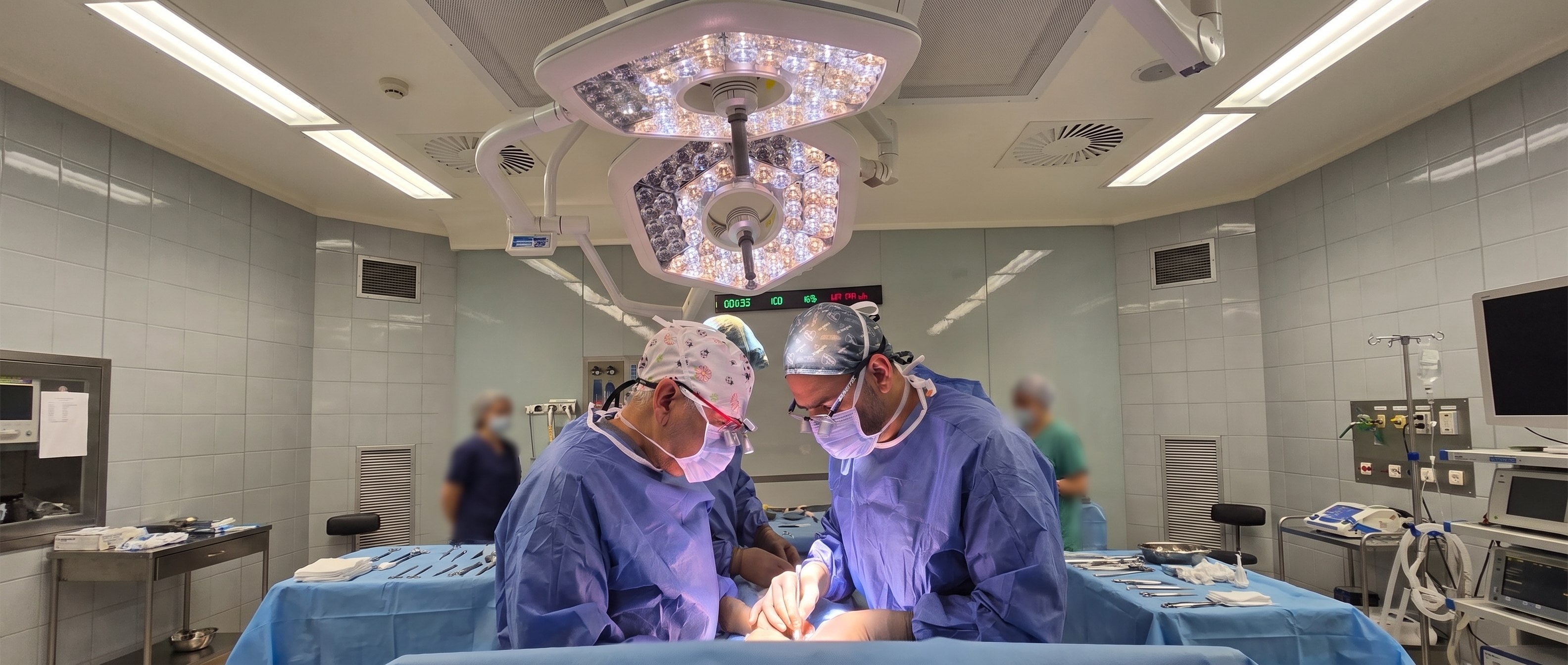
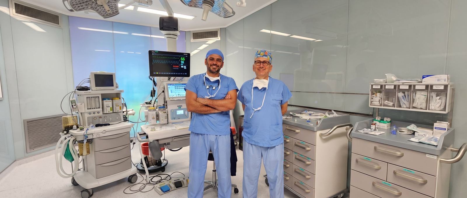
Two pediatric urology specialists work together on every operation: one performs the repair while the other assists, checks, and takes over when needed. This built-in second opinion completes demanding operations with less risk.

  • Lower risk

    A shared assessment and four-handed technique; a second experienced pair of eyes at the demanding steps.

  • Shorter surgery

    Working in parallel shortens the time spent under anesthesia.

  • Less anesthesia

    Infants and young children spend less time under anesthesia.

Prof. Dr. Ali Avanoğlu — hypospadias surgeon, Turkey

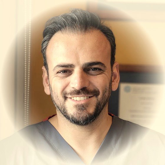
Prof. Dr. Ali Avanoğlu

Pediatric Surgeon and Pediatric Urologist

Over forty years in medicine, thirty-two of them as a pediatric surgeon; focused exclusively on pediatric urology since 1994. Associate professor in 1996 and full professor in 2002 at Ege University, where he chaired both the Department of Pediatric Surgery and the Division of Pediatric Urology. Roughly 3,000 hypospadias repairs and more than 1,000 operations for vesicoureteral reflux.

Doç. Dr. Yaşar Issı — hypospadias surgeon, Turkey

Doç. Dr. Yaşar Issı

Pediatric and Adult Urologist

Graduate of Ege University Faculty of Medicine (2006); completed urology training at İzmir Atatürk Training and Research Hospital (2011) and a pediatric urology fellowship at Ondokuz May University (2016); associate professor since 2022. Has worked at Gaziantep Children's Hospital, İzmir Çiğli Training and Research Hospital, and Bakırçay University. More than 1,000 hypospadias repairs and a comparable number of stone operations; continues the same patients' follow-up from childhood through adolescence and adulthood, in the same team.

Why together?

What operating together means for your child

In every operation, it is not a surgeon and an assisting resident — it is two specialists who both know the procedure, working together. We summarize the difference in four points.

Difference 01

Critical decisions are made together

Which technique to use, whether the repair will finish in one session, and how to respond to anything unexpected during surgery are decided by two specialists' shared assessment — never by one person alone.

Shared decision
2×
Two pediatric urology surgeons
Together, in the same team, every time
Two hypospadias surgeons operating together
Difference 02

Shorter time under anesthesia

While one surgeon finishes a step, the other prepares the next; four hands work at once. In an infant or young child, every extra minute under anesthesia is a risk in its own right.

Two hands, at the same time
Difference 03 Related conditions, same session
1×

One anesthesia: conditions found together (undescended testis, inguinal hernia, hydrocele, hydronephrosis) are planned into the same session.

Get in touch
Difference 04 — Follow-up never breaks off

Some issues only surface during adolescence or adulthood. Because the team includes both pediatric and adult urology, follow-up never breaks off between childhood and adulthood.

Pediatric urology
Adult urology
Same team, same record
Process · for patients travelling from abroad too

Travelling to Turkey for treatment?

Surgery takes place at İzmir Acıbadem Kent Hospital; our clinic is about 4–5 km from the hospital, with a range of accommodation options nearby. The process runs in coordination between the clinic and the hospital, from your first consultation through to the completion of post-operative checks.

Clinic Hospital İzmir Acıbadem Kent Hospital · about 4–5 km.
  1. 01 Clinic
    Pre-treatment review with two pediatric urology surgeons — family on a video call from home

    Consultation and surgical planning

    Patients are first assessed at our clinic; surgical planning is done here.

    Avanoğlu–Issı Clinic

  2. 02 Hospital
    Prof. Dr. Ali Avanoğlu and Doç. Dr. Yaşar Issı, hypospadias surgeons in İzmir, Turkey

    Pre-operative tests and anesthesia review

    One day before surgery, you are referred to İzmir Acıbadem Kent Hospital for the required tests and anesthesia assessment.

    One day before surgery

  3. 03 Hospital
    Two pediatric urology surgeons' treatment process — family arriving at the hospital

    Surgery

    The operation takes place the next day, as planned. In suitable cases, it is completed as day surgery.

    İzmir Acıbadem Kent Hospital

  4. 04 Clinic
    Two pediatric urology surgeons' treatment process — remote follow-up after surgery, mother on a video call from home

    Recovery after discharge

    After the observation period, patients who meet discharge criteria continue recovering at home or at their accommodation. Dressing changes, wound checks, and catheter checks and removal continue at our clinic.

    Avanoğlu–Issı Clinic

Frequently asked by families

What families travelling from abroad ask first

Short answers are below; write to us directly with any question about your trip.

Let's plan your trip together
  • İzmir, Turkey
  • Two pediatric urology specialists
  • Same team from diagnosis to follow-up
Where do the operations take place?
Surgery takes place at İzmir Acıbadem Kent Hospital. Our clinic is about 4–5 km from the hospital, with a range of accommodation options nearby.
How does the process work, from consultation to surgery?
You are first assessed at our clinic, where the surgical plan is made. One day before surgery you are referred to the hospital for the required tests and anesthesia review; the operation itself takes place the following day, as planned.
Do we stay in hospital after surgery?
A significant share of our operations, when suitable, are completed as day surgery. Patients who meet discharge criteria continue recovering, after the observation period, in the more comfortable setting of your home or accommodation.
How are post-discharge checks handled?
Follow-up steps such as dressing changes, wound checks, and catheter checks and removal continue to take place at our clinic. That way, the entire process — from the first consultation through to the completion of follow-up — runs in coordination between the clinic and the hospital.
Topic index • 7 clusters

All vesicoureteral reflux topics

7 clusters, 43 detail pages. Each page focuses on a single question and links back here, to the main treatment page.

Scientific publications · 22 articles

Prof. Dr. Ali Avanoğlu and Doç. Dr. Yaşar Issı's publications on Vesicoureteral Reflux (VUR)

Articles indexed in PubMed; the complete set of citations from the pages above. For all publications, see the Publications page →

  1. Tosun H, Issi Y, Akgul AK, et al. (2026). Is YouTube an Accurate and/or Comprehensive Source of Information on Monosymptomatic Nocturnal Enuresis? Journal of clinical practice and research. PubMed ↗
  2. Tiryaki S, Issi Y (2023). The association of meatal stenosis and infant circumcision. The Turkish journal of pediatrics. PubMed ↗
  3. Tiryaki S, Tekin A, Avanoglu A, et al. (2022). A pilot study assessing average detrusor pressure garnered from area under a urodynamic curve: Evaluation of clinical outcomes. Journal of pediatric urology. PubMed ↗
  4. Tiryaki S, Celtik U, Eraslan C, et al. (2022). Diffusion tensor imaging in anorectal malformations: a pilot study for the evaluation of lumbosacral plexus. Pediatric surgery international. PubMed ↗
  5. Issi Y, Biçakci U (2021). Does desmopressin withdrawal strategy affect relapse rates in monosymptomatic enuresis treatment? European journal of pediatrics. PubMed ↗
  6. Issi Y (2020). Non-Invasive Stent Removal after Ureteroneocystostomy in Pediatric Patients: Long-Term Results. Urology journal. PubMed ↗
  7. Tiryaki S, Eraslan C, Soyer T, et al. (2019). Nonneuropathic Neuropathic Bladder-Is it Really Nonneuropathic? The Journal of urology. PubMed ↗
  8. Tekin A, Yagmur I, Tiryaki S, et al. (2018). Changing bulking agent may require change in injection volume for endoscopic treatment of vesicoureteral reflux. International braz j urol. PubMed ↗
  9. Tiryaki S, Yagmur I, Parlar Y, et al. (2015). Botulinum injection is useless on fibrotic neuropathic bladders. Journal of pediatric urology. PubMed ↗
  10. Kocherov S, Ulman I, Nikolaev S, et al. (2014). Multicenter survey of endoscopic treatment of vesicoureteral reflux using polyacrylate-polyalcohol bulking copolymer (Vantris). Urology. PubMed ↗
  11. Tiryaki S, Alkac AY, Serdaroglu E, et al. (2013). Involution of multicystic dysplastic kidney: is it predictable? Journal of pediatric urology. PubMed ↗
  12. Tiryaki S, Ergun O, Celik A, et al. (2010). Success of Malone's antegrade continence enema (MACE) from the patients' perspective. European journal of pediatric surgery. PubMed ↗
  13. Ural Z, Ulman I, Avanoglu A (2008). Bladder dynamics and vesicoureteral reflux: factors associated with idiopathic lower urinary tract dysfunction in children. The Journal of urology. PubMed ↗
  14. Ozcan C, Ergün R, Ozbek SS, et al. (2007). Bladder ultrasound in the evaluation of the efficacy of dextranomer/hyaluronic acid injection for treatment of vesicoureteral reflux. Journal of clinical ultrasound. PubMed ↗
  15. Ozel SK, Dokumcu Z, Akyildiz C, et al. (2007). Factors affecting renal scar development in children with spina bifida. Urologia internationalis. PubMed ↗
  16. Sözübir S, Ergun G, Celik A, et al. (2006). The influence of urine osmolality and other easily detected parameters on the response to desmopressin in the management of monosymptomatic nocturnal enuresis in children. Minerva urologica e nefrologica. PubMed ↗
  17. Sözübir S, Celik A, Emir N, et al. (2005). Consistency of urodynamic parameters in children with detrusor instability: how many times should the bladder be filled? Urologia internationalis. PubMed ↗
  18. Ergun O, Celik A, Corduk N, et al. (2003). De-inversion of an inverted appendectomy for the Malone procedure. BJU international. PubMed ↗
  19. Celik A, Ulman I, Aydin M, et al. (2002). Familial vesicoureteral reflux in asymptomatic siblings. The Turkish journal of pediatrics. PubMed ↗
  20. Ulman I, Avanoğlu A, Erikçi V, et al. (1998). Is resolution of vesicoureteric reflux by conservative management predictable in patients with myelodysplasia? European urology. PubMed ↗
  21. Ulman I, Avanoğlu A, Erdener A, et al. (1996). Evaluation of the lower urinary tract function in caudal duplication (dipygus) anomaly. Journal of pediatric surgery. PubMed ↗
  22. Ulman I, Avanoğlu A, Gökdemir A (1995). Neuroanatomy of the human striated urethral sphincter. British journal of urology. PubMed ↗
Contact us

With the vesicoureteral reflux assessment that fits your situation would you like to reach the doctor?

There are two ways. If you'd like the doctor to already know your situation before replying, start with the short assessment; if your question is brief, write to us directly. Both reach the same team.

1 Let me assess my situation firstRecommended The Roadmap asks a few questions in about 2 minutes, collects your answers with an anatomical diagram, and produces a ready-made summary. That summary is added to your WhatsApp message — the doctor replies already knowing your situation. Start the Roadmap
2 I'll write directly Fill in the form; your message opens ready-made in WhatsApp (you can edit it before sending), or you can send it directly by email. This page's topic is added to the message automatically.

Editor's note: Age and a short sentence are enough; you don't need to use medical terms. Please don't send photos — the doctor will request them through a secure channel if needed. What you write is only sent when you submit it; this page does not store anything.

Direct contactReply comes from the doctor
Who is this for?
Quick questions:

Your message is not stored on our server; it is sent by WhatsApp/email. Privacy notice →

Gomauna Web Design and Development gomauna.com Prof. Dr. Ali Avanoğlu | 2026 © All Rights Reserved. SEO & GEO OPTIMIZED
WhatsApp Roadmap