
Skip to content
Prof. Dr. Ali Avanoğlu
Main treatment page • Undescended Testicle

Undescended Testicle Surgery in Turkey

An undescended testicle is one that has not descended into the scrotum at birth, or that does not remain there afterward. It is one of the most common congenital urological differences in baby boys. Treatment is not a single named operation: it is a set of pathways that vary according to where the testicle is, whether it can be felt, and the child's age. This page walks through that whole picture.

ByProf. Dr. Ali AvanoğluUpdated
At a glance 42 pages • 15 video
6–12 months
Surgical age

The guideline target; completed by around 18 months at the latest

Usually one
Number of operations

A planned two-stage approach for a high intra-abdominal testicle

Two specialists
Surgical team

Examination, laparoscopy and repair, all with the same team

The whole treatment • 12 sections

This page explains the whole picture; the details of each topic are in the pages below

Start with what an undescended testicle is and how the testicle descends; then diagnosis, types, palpable and non-palpable testicles, surgical timing, techniques, the staged approach, after surgery, the adolescent–adult period, and follow-up: twelve sections. Each section has detail pages below it; jump to the one you want from the contents on the right (at the top on mobile).

undescended testicle
01Section 1 / 12

What is an undescended testicle?

Usually noticed at the very first exam

An undescended testicle (cryptorchidism, in medical terms) is a testicle that is not found inside the scrotum at birth, or that does not stay there even if it is brought down. The testicle normally forms near the kidneys before birth and descends through the inguinal canal into the scrotum in the last months of pregnancy; when this journey is not completed, the testicle remains inside the abdomen, in the inguinal canal, or just above the scrotum.

IN THE INGUINAL CANALRIGHTLEFT
Example: the left testicle has remained in the inguinal canal; the dashed line shows the descent path, the ghost outline shows the target position in the scrotum
Definitionthree findings
  • 1Scrotum empty or smallthe testicle is not in the scrotum: inside the abdomen, in the inguinal canal, or just above the scrotum
  • 2No painfamilies usually notice by appearance; the baby is not bothered
  • 3Diagnosis is made by examinationtwo questions: can it be felt, and where?
Quick factsat a glance
Frequency1–4% in full-term babies; up to 30% in premature babies
BilateralRoughly one-third of cases
Spontaneous descentFirst 6 months; not expected after that
TreatmentOrchiopexy; target 6–12 months
SurgeryUsually a single operation; a planned two-stage approach for a high intra-abdominal testicle
Not to be confused with a retractile testicle

It descends into the scrotum and stays there; it is not a disease and does not require surgery — but it is monitored until puberty.

Read the full text
  • It occurs in roughly 1–4% of full-term baby boys, and at a much higher rate in premature babies — up to 30%. About one-third of cases are bilateral. In some babies the testicle descends on its own in the first months after birth; after six months, this becomes much less likely.
  • An undescended testicle does not cause pain and does not bother the baby; families usually notice it because the scrotum looks empty or small on one side. The diagnosis is made by examination — determining where the testicle is and whether it can be felt is where all planning begins.
  • The condition most often confused with it is the 'retractile testicle': the testicle is in the scrotum, but it draws upward when it contracts in the cold or when touched, and stays in the scrotum once it is guided back down by hand. This is not a disease and usually does not require surgery — but it does need monitoring, since in some children it can genuinely move upward over time.
Why does it matter?

The scrotum keeps the testicle a few degrees cooler than the rest of the body, which is the environment it is built to work in. When it stays inside the abdomen or the groin, the sperm-producing cells are damaged over time. Treatment is not done for appearance — it is done to protect future fertility potential and to keep the testicle somewhere it can be examined.


Videos in this sectionFull guide (15) →
How does the testicle descend, and why doesn't it?
02Section 2 / 12

How does the testicle descend, and why doesn't it?

The testicle descends into the scrotum in the last months of pregnancy

The testicle forms inside the abdomen, near the kidney, in the early weeks of pregnancy. From the middle of pregnancy onward it moves downward, and in the last three months it passes through the inguinal canal and settles into the scrotum. This descent is a process in which hormones (testosterone in particular, along with some signals the testicle itself produces), a guiding band of connective tissue called the gubernaculum, and abdominal pressure all work together.

Descentthree steps
  1. 1Inside the abdomen — first half of pregnancythe testicle forms near the kidney
  2. 2Into the inguinal canal — after mid-pregnancyguided to the internal ring by the gubernaculum and hormonal signals
  3. 3Into the scrotum — the last three monthspasses through the inguinal canal and settles into the scrotum
Contributing factorstaken together
  • 1Premature birth, low birth weightthe most strongly associated factors
  • 2Family tendencya history in the father or a sibling
  • 3Hormonal signalscan fall short; most cases have no single identifiable cause
  • 4Ascending testiclein the scrotum as a baby, moves upward while growing
Read the full text
  • In premature babies, the descent has not yet finished, which is why undescended testicles are much more common in this group; in some of these babies, the testicle still comes down on its own in the first months after birth. In full-term babies, though, a testicle that is still undescended at six months is no longer expected to descend by itself.
  • There is no single cause. Premature birth and low birth weight are the most strongly associated factors; a family history of undescended testicle, insufficient hormonal signaling, and certain pregnancy-related factors can also play a role. In most cases, no single identifiable cause is found.
  • The testicle is not always high from birth. In some children, the testicle is in the scrotum as an infant, but the spermatic cord fails to lengthen along with the child's growth, and the testicle moves upward over the years; this is called an 'ascending testicle.' So the fact that 'it was in the scrotum as a baby' does not, by itself, mean everything is fine — the testicle's position is checked at exams up through school age.
Two questions families often ask

Did we do something wrong? No — it is not the result of anything done, or not done, during pregnancy. Will it happen again? There can be a family tendency, but in most families it occurs in only one child.

Videos in this sectionFull guide (15) →
undescended testicle in babies
03Section 3 / 12

How is an undescended testicle recognized? Examination and diagnosis

Diagnosis is made by examination, not imaging

The diagnosis and classification of an undescended testicle are made by examination; imaging does not replace this. An experienced examination answers two basic questions: can the testicle be felt, and if so, where? These two answers largely determine the treatment pathway.

Examinationstep by step
  1. 1A warm room, a calm childthe cremasteric reflex is minimized
  2. 2The inguinal canal is stroked from top to bottomtrying to guide the testicle toward the scrotum
  3. 3Does it stay once released?if it stays, it's retractile; if it springs back up immediately or never comes down, it's a true undescended testicle
  4. 4If it can't be felteither inside the abdomen or absent → examination under anesthesia, and diagnostic laparoscopy if needed
Imagingwhen it's actually used
MethodWhat it showsIts role
UltrasoundCan show a testicle in the groin; not reliable for inside the abdomen — 'not seen' does not mean 'absent'Not routinely recommended
MRILimited sensitivity; may require sedationNot routinely recommended
Diagnostic laparoscopyDirect view inside the abdomen; treatment can proceed in the same sessionThe standard for a non-palpable testicle
Read the full text
  • The examination is done in a warm room, with the child calm. The examiner strokes down along the inguinal canal, trying to guide the testicle toward the scrotum. If the testicle comes down and stays there once released, it is a retractile testicle; if it comes down but immediately springs back up when released, or does not come down at all, it is a true undescended testicle.
  • If the testicle can be felt in the groin, its position, size, and how it compares with the other testicle are noted. If it cannot be felt, there are two possibilities: the testicle is inside the abdomen, or it is absent / never developed. Non-palpable testicles make up roughly one-fifth of cases, and some of these are indeed inside the abdomen.
  • Ultrasound and MRI do not reliably show the location of a non-palpable testicle; not seeing it on imaging does not mean it is absent, and seeing it does not always give the correct location either. For this reason, guidelines do not routinely recommend imaging for a non-palpable testicle; the definitive evaluation is a repeat examination under anesthesia and, if needed, diagnostic laparoscopy.
  • Bilateral non-palpable testicles are a separate topic: they call for hormonal, and if needed, genetic evaluation; when an additional finding such as hypospadias is present, pediatric urology, endocrinology, and genetics work together.
Sudden, severe pain is an emergency

In a child with an undescended or ascending testicle, sudden, severe pain in the groin, abdomen, or testicle needs urgent evaluation for testicular torsion. Do not wait — go to the nearest emergency department.

Videos in this sectionFull guide (15) →
Prof. Dr. Ali Avanoğlu's publications on this topic · 1
  1. Ulman I, Sakalli U, Avanoğlu A, et al. (1996). Serum creatine kinase enzyme levels in the early diagnosis of spermatic cord torsion. Urological research. PubMed ↗
what is a retractile testicle
04Section 4 / 12

Types of undescended testicle: retractile, ascending, true undescended, ectopic

The same picture can mean four different things

What looks like the same picture can actually correspond to four different situations, and each one follows a different pathway. What tells them apart is how the testicle behaves on examination, together with the history.

RIGHTLEFT
Retractile
RIGHTLEFT
Ascending
RIGHTLEFT
True undescended
RIGHTLEFT
Ectopic
Four patterns: retractile (oscillates), ascending, true undescended, ectopic
Four pictureshow they behave on examination
TypeOn examinationPathway
Retractiledescends into the scrotum and stays; pulled up from time to timeno surgery; checked once a year until puberty
Ascendingwas in the scrotum as a baby, moved up while growingorchiopexy is usually planned
True undescendedstopped along the path of descent; cannot be brought down, or doesn't stayorchiopexy; more extensive the higher it sits
Ectopicoutside the normal path: superficial inguinal pouch, base of the thigh, perineum, base of the penissurgical repositioning
Read the full text
  • Retractile testicle: the testicle descends into the scrotum and stays there for a while once released; it is pulled up from time to time by the cremasteric reflex. It does not require surgery, but it is checked regularly — at least once a year — until puberty, because some retractile testicles turn into ascending testicles over time.
  • Ascending testicle: a testicle known to have been in the scrotum in infancy moves upward as the child grows. It is usually noticed in the preschool or school-age years. It is assessed by pediatric urology, and orchiopexy — fixing the testicle in the scrotum — is usually planned.
  • True undescended testicle: the testicle has stopped somewhere along its normal path of descent — inside the abdomen, in the inguinal canal, or just above the scrotum — and cannot be brought down into the scrotum by hand, or does not stay there if it is. The higher its position, the more extensive the surgery.
  • Ectopic testicle: the testicle has completed its descent but has ended up in the wrong place — such as the superficial inguinal pouch, the base of the thigh, the perineum, or the base of the penis. It cannot be brought into the scrotum by hand; treatment means surgically repositioning it.
Videos in this sectionFull guide (15) →
Prof. Dr. Ali Avanoğlu's publications on this topic · 1
  1. Divarcı E, Ulman I, Avanoglu A (2011). Transverse testicular ectopia treated by transseptal contralateral transposition: case report. European journal of pediatric surgery. PubMed ↗
Palpable undescended testicle: in the groin or in a low position
05Section 5 / 12

Palpable undescended testicle: in the groin or in a low position

The large majority of cases; the most predictable group to treat

An undescended testicle that can be felt on examination accounts for the large majority of cases, and it is the group with the most predictable treatment. The testicle is most often in the inguinal canal; in some children it sits at the canal's exit, in a 'low' position close to the scrotum.

RIGHTLEFT
In the inguinal canal
RIGHTLEFT
Low-lying
RIGHTLEFT
Ectopic
Three locations of a palpable testicle
Two approachesfor a palpable testicle
Inguinal orchiopexya testicle in the inguinal canal
  • Small incision in the groin
  • A hernia sac, if present, is closed in the same session
  • The standard approach; day surgery
Scrotal orchiopexylow-lying, easily brought down
  • Single incision through the scrotum
  • No groin incision needed
  • Selected cases; faster recovery
The processin brief
AnesthesiaGeneral
Duration30–60 minutes, one-sided
Hospital stayUsually day surgery
DecisionMade on examination and under anesthesia
Read the full text
  • For a testicle in the inguinal canal, the standard operation is inguinal orchiopexy: through a small incision in the groin, the testicle and its vessels are freed from the surrounding tissue, any accompanying hernia sac is closed, and the testicle is brought down into the scrotum without tension and fixed in place. This is usually completed in a single operation, as day surgery.
  • For testicles that sit close to the scrotum, in a low position, and can be brought down easily, repair through a single incision in the scrotum alone (scrotal orchiopexy) is possible in selected cases; no groin incision is needed, and recovery is faster. Which approach is appropriate is decided on examination and under anesthesia.
  • The ectopic testicle also belongs to the palpable group; the difference is that the testicle lies outside its normal path. Treatment is still surgical placement into the scrotum, with the incision planned according to where the testicle is found.
How long does the operation take?

A one-sided inguinal or scrotal orchiopexy usually takes 30–60 minutes, is done under general anesthesia, and the child usually goes home the same day.

Videos in this sectionFull guide (15) →
cryptorchidism
06Section 6 / 12

Non-palpable testicle: intra-abdominal, peeping and vanishing testis

The evaluation is completed in the operating room

A testicle that cannot be felt on examination makes up roughly one-fifth of cases, and planning proceeds differently here. The first question is whether the testicle exists at all; the second is where it is. Ultrasound and MRI do not answer these questions reliably, so the evaluation is carried out in the operating room.

RIGHTLEFT
Peeping
RIGHTLEFT
Inside the abdomen
RIGHTLEFT
High intra-abdominal
?RIGHTLEFT
Vanishing / absent
Four findings that may be seen on laparoscopy
In the operating roomthe sequence
  1. 1Examination under anesthesiasome testicles that can't be felt while awake can be felt once the child is asleep → standard orchiopexy
  2. 2Diagnostic laparoscopya thin camera through the navel; the abdomen is seen directly
  3. 3Decision based on findingsin the same session: bringing it down, opening the groin, a staged approach, or 'no testicle to bring down'
What laparoscopy can showwhat's done
FindingWhat it meansNext step
Testicle near the internal ring (peeping)can be brought downsingle-stage orchiopexy
Inside the abdomen, vessels adequatecan be brought downlaparoscopic orchiopexy
Very high, vessels shortforcing it down would disrupt blood supplyFowler–Stephens or Shehata (staged)
Vessels enter the canaltesticle or a remnant is in the groininguinal exploration
Vessels end blindlyvanishing testisno further surgery needed
Read the full text
  • The first step is a repeat examination under anesthesia: some testicles that cannot be felt while the child is awake, because of muscle contraction, can be felt once the child is asleep, and the case is completed with a standard orchiopexy. If it still cannot be felt, diagnostic laparoscopy is performed — a thin camera inserted through the navel is used to look inside the abdomen.
  • Laparoscopy can reveal several different pictures. If the testicle is inside the abdomen and its vessels are long enough, it is brought down into the scrotum in the same session with laparoscopic orchiopexy. If the testicle sits just next to the internal ring (a 'peeping' testicle), orchiopexy — laparoscopic or through the groin — is usually possible in a single stage.
  • If the vessels enter the inguinal canal, the groin is opened (inguinal exploration): if a viable testicle is found, it is brought down into the scrotum; if only a small, non-functioning remnant (a nubbin) is found, it is removed. If the vessels are seen to end blindly inside the abdomen, the testicle was lost before birth due to a circulation problem — a 'vanishing testis' — and in that case there is no testicle to bring down, and no further surgery is needed.
  • If the testicle is very high, with vessels too short to reach the scrotum, bringing it down in a single session would put its blood supply at risk; in these cases, a planned staged approach — Fowler–Stephens or Shehata — comes into consideration. This decision is made during laparoscopy, which is why this possibility needs to be discussed with the family beforehand.
Why does the other testicle get bigger?

In a one-sided non-palpable testicle, a noticeably enlarged testicle on the other side can suggest it has taken over the function of the missing one — but this finding alone does not confirm the diagnosis, and it does not remove the need for laparoscopy.

Videos in this sectionFull guide (15) →
undescended testicle surgery
07Section 7 / 12

When should undescended testicle surgery be done?

Target 6–12 months; 18 months at the latest

Timing is one of the most important factors determining the outcome in an undescended testicle. In the first six months after birth, the testicle may still descend on its own, and this period is followed with examinations. Once six months is reached (using corrected age for babies born prematurely), spontaneous descent is no longer expected for a testicle that is still undescended, and surgical planning begins.

OBSERVATIONCan descend on its ownTARGET WINDOWOrchiopexyAT THE LATESTDon't delayFOLLOW-UP · DECISIONEvaluated at any ageBIRTH6 MO12 MO18 MOPUBERTYADULTRECOMMENDED WINDOW FOR SURGERY
0–6 months observation · 6–12 months target · ≈18 months at the latest · evaluation in adolescents and adults
Timelineby age
0–6 monthsMonitoring; it may still descend on its own
6 monthsIf still undescended, surgical planning begins (corrected age for premature babies)
6–12 monthsThe target surgical window
≈18 monthsCompletion at the latest
After thatAscending testicle, adolescents, adults: evaluated at any age
Why early?two reasons
  • 1Fertility potentialthe precursor cells for sperm decline over the years in a warm environment
  • 2Cancer risk and examinationorchiopexy before puberty lowers the risk; a testicle in the scrotum is easy to examine
Hormone treatment

Not routinely recommended to bring down a one-sided testicle; considered together with endocrinology in selected bilateral cases.

Read the full text
  • Current guidelines recommend orchiopexy between 6 and 12 months of age, completed by around 18 months at the latest. The reason for this window lies in the tissue itself: the precursor cells that go on to make sperm decline over the years the longer the testicle stays in a warm environment; doing the surgery early limits this loss and helps protect future fertility potential.
  • The second reason for early surgery is testicular cancer risk. People with a history of an undescended testicle have a higher risk of testicular cancer than the general population; orchiopexy performed before puberty has been reported to lower this risk, while the risk stays notably higher when repair is done late, or not at all. A testicle in the scrotum is also easy to examine.
  • For cases recognized late — an ascending testicle, or a case noticed in adolescence or adulthood — treatment is never abandoned on the thought that 'it's too late'; evaluation is carried out at any age, and the decision is made individually, based on the testicle's condition, the other testicle, and the patient's priorities.
  • Hormone treatment (hCG or GnRH) is not routinely recommended to bring a one-sided undescended testicle down; the success rate is low, and the testicle often moves back up afterward. In bilateral cases, endocrine treatment can be considered in selected situations to support fertility potential; this decision is made together with pediatric urology and endocrinology.
Quick summary

0–6 months: monitoring (it may still descend on its own). After 6 months: if still undescended, surgical planning begins. Target window: 6–12 months; completed by ~18 months at the latest. Evaluation is also carried out in adolescents and adults.

Videos in this sectionFull guide (15) →
Undescended testicle surgery: approaches and techniques
08Section 8 / 12

Undescended testicle surgery: approaches and techniques

The shared name is orchiopexy; the entry point and number of stages vary

The common name for all of these approaches is orchiopexy: freeing the testicle and its vessels from the surrounding attachments, then bringing it down into the scrotum without tension and fixing it in place. What changes, depending on where the testicle is, is the surgical entry point and the number of stages needed to complete it.

Approachesby the testicle's position
MethodWhat's doneWhen
Inguinal orchiopexygroin incision; the testicle is freed and fixed in the scrotum; hernia sac closeda testicle palpable in the groin
Scrotal orchiopexysingle incision through the scrotumlow-lying, selected cases
Laparoscopic orchiopexyminimally invasive; diagnosis and treatment in the same sessionan intra-abdominal testicle
Fowler–Stephens (two stages)vessel divided → ≈6 months → bringing it downhigh intra-abdominal
Shehata (traction)fixed under traction → ≈12 weeks → bringing it downhigh intra-abdominal
Redo orchiopexyre-mobilization within scar tissuea testicle that has moved back up
Orchiectomyremoval of the testicleselected cases: non-functioning / atrophic; adolescents–adults
Read the full text
  • Inguinal orchiopexy is the standard approach for a testicle that can be felt in the groin; an accompanying hernia sac is closed in the same session. Scrotal orchiopexy is done through a single incision in the scrotum, for selected, low-lying testicles. Laparoscopic orchiopexy is used for a testicle inside the abdomen; both diagnosis and treatment can be carried out in the same session.
  • For a high intra-abdominal testicle, when the vessels are not long enough to reach the scrotum, two-stage methods come into play. In the Fowler–Stephens technique, the main testicular vessel is divided, and the testicle is then nourished by collateral circulation running alongside the vas deferens; roughly six months after the vessel is divided, the testicle is brought down into the scrotum in a second session. In the Shehata (traction) technique, the vessel is not divided: in the first session, the testicle is fixed to the inner surface of the abdominal wall, stretched toward the opposite side; the vessels are then given about 12 weeks to lengthen before the testicle is brought down into the scrotum in a second session.
  • In children who have had previous surgery and whose testicle has moved back up, redo orchiopexy is performed; because of scar tissue, it is planned even more carefully than the first operation. For a high-lying testicle, or one thought to be small and non-functioning, removing it (orchiectomy) is also discussed as an option in certain selected cases; this decision is made individually, particularly in adolescents and adults.
  • Which approach will be used can largely be anticipated before surgery, but for a non-palpable testicle the final decision only becomes clear during the operation, based on what laparoscopy shows. This is why, during the consent discussion, the goal of a single session and the possibility of a staged approach are both discussed together.
Videos in this sectionFull guide (15) →
High intra-abdominal testicle: the staged approach
09Section 9 / 12

High intra-abdominal testicle: the staged approach

The staged approach: a strategy planned from the start to protect the testicle

Some intra-abdominal testicles sit close to the internal ring and can be brought down in a single session. When the testicle is higher and its vessels stay short, two risks come into conflict: forcing it down stretches the vessels and disrupts its blood supply, while not bringing it down leaves the testicle in a warm environment. Staged techniques were developed to resolve this dilemma.

STAGE 1Main testicular vesseldivided / ligated≈ 6 MONTHSCollateral circulationstrengthensSTAGE 2Testicle broughtinto the scrotum
STAGE 1Testicle fixed toabdominal wall under traction≈ 12 WEEKSVesselslengthenSTAGE 2Testicle broughtinto the scrotum
Top: Fowler–Stephens (vessel divided, ≈6 months); bottom: Shehata (fixed under traction, ≈12 weeks)
Two techniquesthe staged approach
Fowler–Stephensthe main vessel is divided
  • Session 1: laparoscopic vessel division
  • ≈6 months: collateral circulation strengthens
  • Session 2: bringing it down into the scrotum
  • Two stages are safer than one
Shehata (traction)the vessel is preserved
  • Session 1: fixed to the abdominal wall under traction
  • ≈12 weeks: the vessels lengthen
  • Session 2: bringing it down into the scrotum
  • Atrophy risk may be lower
The staged approach is not a failure

It is a strategy planned from the start to protect the testicle; the child continues normal life in between.

Read the full text
  • In the Fowler–Stephens approach, the main testicular vessel is divided or ligated laparoscopically; the testicle then continues to be nourished by the collateral vessels running along the vas deferens. About six months are allowed for the collateral circulation to strengthen, and the testicle is brought down into the scrotum in a second session. When Fowler–Stephens is done in a single session, the risk of losing the testicle is higher; with the two-stage approach, the reported rate of testicular survival in the literature is notably better.
  • In the Shehata technique, the vessel is not divided. In the first session, the testicle is fixed laparoscopically to the inner surface of the abdominal wall, at a point on the opposite side, so that the vessels are gradually stretched and lengthen over time. About twelve weeks later, the testicle is placed into the scrotum in a second session. Because the main vessel is preserved, the risk of atrophy has been reported to potentially be lower; which technique is chosen depends on how high the testicle sits and on the vessel anatomy.
  • With either technique, the child continues normal life in between the two sessions. The most important thing for families to understand is that the staged approach is not a failure — it is a strategy planned from the start specifically to protect the testicle.
  • Even after a staged operation successfully brings the testicle into the scrotum, its size may remain smaller than the other testicle, and the need for long-term follow-up is discussed from the outset.
Why is 'two stages' discussed from the start?

For a non-palpable testicle, the true picture is only seen at laparoscopy. During the consent discussion, both the goal of a single-session repair and the possibility of switching to a staged approach are explained together; the decision is made during the same session, based on what is found.

Videos in this sectionFull guide (15) →
undescended testicle surgery recovery in babies
10Section 10 / 12

After undescended testicle surgery: care and possible issues

Usually day surgery; swelling is expected in the first few days

Orchiopexy is usually a day-surgery procedure; the child goes home the same day. Swelling, bruising, and tenderness in the groin and scrotum are expected in the first few days; pain is controlled with simple pain relievers. The wound is kept covered, and bathing starts after the interval the doctor specifies; activities that put pressure on the area, such as horseback riding or cycling, are avoided for a few weeks.

At homethe first weeks
The first daysswelling, bruising, tenderness; simple pain relievers
The woundkept covered; bathing starts after the interval the doctor specifies
Activityno pressure-bearing activities such as cycling or horseback riding for a few weeks
Follow-up checkthe testicle's position, size, and the wound
Watched over the long termpossible issues
  • 1Ascending againpulled back toward the groin by adhesions → redo evaluation
  • 2Atrophyshrinkage if blood supply is compromised; risk rises with a higher starting position
  • 3Herniaa new swelling in the groin
  • 4Painpersisting or newly starting
Read the full text
  • At the postoperative check, the testicle's position in the scrotum, its size, and the wound are assessed. Wound infection or bleeding are occasionally seen early on, though rarely; fever, increasing redness, and discharge should be reported to the doctor.
  • Two things are watched over the long term. The first is the testicle ascending again: it can be pulled back from the scrotum toward the groin due to adhesions or incomplete mobilization during surgery; in that case, redo orchiopexy is considered. The second is testicular atrophy: the testicle can shrink if the vessels were stressed during surgery, or if the blood supply was insufficient in a staged procedure; the risk is higher the higher the testicle started out.
  • A new swelling in the groin is evaluated for a possible hernia, and pain that persists or starts newly after surgery is also assessed separately. Most of these issues can be resolved if caught early, which is why regular follow-up matters.
When to contact the doctor

Fever; increasing redness or discharge at the wound; bleeding that does not stop; noticeable and increasing swelling in the scrotum; sudden, severe pain.

Videos in this sectionFull guide (15) →
undescended testicle diagnosed late
11Section 11 / 12

Undescended testicle in adolescents and adults

In adolescents and adults, the decision is individual

An undescended testicle is not always noticed in infancy; some cases come to light in adolescence or adulthood, through an examination, an infertility work-up, or incidental imaging. At these ages, decisions are made on different principles than in children: the goal is not simply to bring the testicle down, but to weigh together the testicle's function, the condition of the other testicle, the wish to have children, and cancer risk.

By agethe decision
StageAssessmentOptions
Adolescentpalpable → orchiopexy; intra-abdominal → the testicle's structure and the other testicleorchiopexy
Adultsize, viability, the other testicle, wish to have children, cancer riskorchiopexy, or orchiectomy in selected cases
Adult operated on in childhoodfertility, shrinkage, a mass, ascending againa separate evaluation pathway for each
Fertility and cancerwhat to know
  • 1One-sidedpaternity rate close to the general population
  • 2Two-sidedsperm parameters and paternity rate drop notably
  • 3Cancer riskhigher than the general population; highest for an intra-abdominal testicle; early orchiopexy lowers it but doesn't eliminate it
  • 4Self-examinationmonthly, from puberty onward
Read the full text
  • In adolescence, if an untreated testicle can be felt on examination, orchiopexy is considered; if it is inside the abdomen, the testicle's structure and the other testicle are considered together. In adulthood, there are two options: bringing the testicle down into the scrotum (orchiopexy), or, in selected cases, removing it (orchiectomy). The decision is made individually, based on age, the size and viability of the testicle, the health of the other testicle, the wish to have children, and cancer risk assessment.
  • For fertility, a one-sided history is very different from a two-sided one. People with a one-sided history have a paternity rate close to that of the general population; with a two-sided history, sperm parameters and the paternity rate are both notably lower. This is why early surgery, and endocrine support when needed, matter even more in bilateral cases.
  • Testicular cancer risk is higher in people with a history of an undescended testicle than in the general population, and it is highest for testicles that remained inside the abdomen. Orchiopexy performed before puberty lowers this risk but does not eliminate it, which is why monthly testicular self-examination is taught from puberty onward, and any suspicious firmness or mass should be evaluated without delay.
  • An adult who had surgery in childhood may come in today because of fertility concerns, a shrinking testicle, a mass, or the testicle being high again; there is a separate evaluation pathway for each of these, and in this team, pediatric urology and adult urology work side by side.
It is not 'too late' after puberty

Evaluation is carried out in adolescents and adults as well; the goal shifts — function, fertility, cancer surveillance — but the decision is individual, and in most cases there is something that can be done.

Videos in this sectionFull guide (15) →
having children after testicle surgery
12Section 12 / 12

Follow-up for an undescended testicle: what changes after surgery?

Follow-up continues from pediatric to adult urology

An undescended testicle is not a case that closes once surgery is done. Whether the testicle that was brought into the scrotum grows, stays in place, and develops as expected at puberty is followed over the years; follow-up continues seamlessly from pediatric urology into adult urology.

Timelinefrom infancy to adulthood
First yearThe testicle's position and size are checked several times
ChildhoodIts place in the scrotum confirmed at yearly exams; also yearly for a retractile testicle
PubertySize and development; hormone ± semen analysis for a testicle that stays small; self-examination is taught
AdultPlans to have children, shrinkage/firmness, swelling in the groin; uninterrupted follow-up with the same team
Read the full text
  • In the first year after surgery, the testicle's position and size are checked several times; after that, its place in the scrotum is confirmed at yearly exams throughout childhood. The same principle applies to monitoring a retractile testicle: at least one examination a year until puberty, because some of them move upward over time.
  • Puberty is a critical period. Testicular size and development are assessed; a testicle that remains noticeably smaller than the other one is evaluated with hormone testing and, if needed, semen analysis. Teenagers are taught testicular self-examination during this period; because early orchiopexy lowers cancer risk without eliminating it, lifelong awareness is essential.
  • In adulthood, the reasons for a visit change: plans to have children, a testicle that has shrunk or feels firm, or swelling in the groin. Having the case continue with the same team from childhood makes it possible to catch late problems early.
Videos in this sectionFull guide (15) →
Roadmap • Interactive tool

Assess your situation yourself

Answer a few questions; the anatomical diagram updates as you go. At the end you're guided to the section and pages that fit your situation, and you can send the summary to the doctor by WhatsApp. About 2 minutes; your answers stay on your device.

Undescended Testicle RoadmapStep-by-step assessment
Step 1
Loading tool…
Undescended Testicle video guide • 15 video

What people ask about Undescended Testicle, in short videos

Prepared in order: first, what an undescended testicle is, the testicle's development and diagnosis; then surgical timing and approaches; and finally follow-up, the adolescent–adult period, fertility, and cancer risk.

Viewing order1–4 / 15
Frequently asked questions

The first 8 questions families ask

Short answers to the questions families ask most often.

Ask your question on WhatsApp
Does an undescended testicle descend on its own?
It can descend in the first six months, and this period is followed with examinations. Once six months is reached (using corrected age for premature babies), if it is still undescended, spontaneous descent is no longer expected, and surgery is planned.
What is the best age for surgery?
Guidelines recommend 6 to 12 months of age, completed by around 18 months at the latest. Cases recognized later are also evaluated, at any age.
Can it be brought down with medication (hormones)?
Hormone treatment is not routinely recommended to bring down a one-sided undescended testicle; success is low, and the testicle often moves back up. In bilateral cases, endocrine treatment can be considered in selected situations to support fertility potential.
We had an ultrasound and the testicle wasn't visible — does that mean it isn't there?
Not seeing it on ultrasound does not mean the testicle is absent; imaging is not reliable for a non-palpable testicle. The definitive evaluation is an examination under anesthesia and, if needed, diagnostic laparoscopy.
Does a retractile testicle need surgery?
Usually not. It is still examined at least once a year until puberty, though, because some retractile testicles genuinely move upward over time (becoming an ascending testicle), and surgery comes into consideration at that point.
How long does surgery take, and will we need to stay in the hospital?
For a one-sided palpable testicle, surgery usually takes 30–60 minutes and is done as day surgery. Laparoscopic and staged operations can take longer; whether a hospital stay is needed depends on the extent of the surgery.
Does an undescended testicle cause infertility?
In people with a one-sided history, the paternity rate is close to that of the general population. With a two-sided history, fertility potential is noticeably reduced, which is why early surgery, and endocrine support when needed, matter. A definitive assessment in adulthood is made with semen analysis and a hormone profile.
Who performs the surgery?
The examination, laparoscopy when needed, and orchiopexy are all carried out by the same team — two physicians, both pediatric urology specialists; follow-up through adolescence and adulthood continues with the same team.
Our surgeons · two specialists, one team

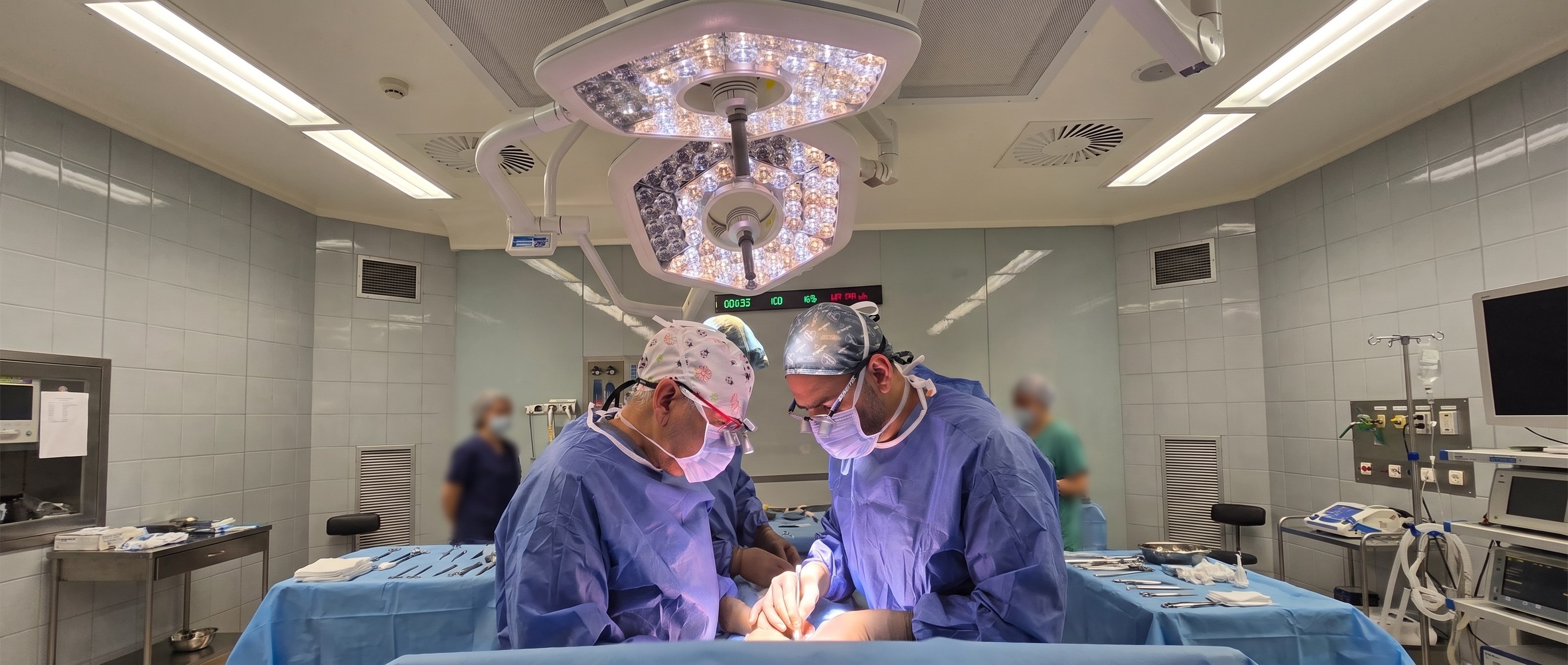
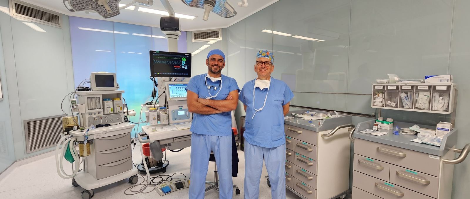
One operation, two surgeons

Two pediatric urology specialists work together on every operation: one performs the repair while the other assists, checks, and takes over when needed. This built-in second opinion completes demanding operations with less risk.

  • Lower risk

    A shared assessment and four-handed technique; a second experienced pair of eyes at the demanding steps.

  • Shorter surgery

    Working in parallel shortens the time spent under anesthesia.

  • Less anesthesia

    Infants and young children spend less time under anesthesia.

Prof. Dr. Ali Avanoğlu — hypospadias surgeon, Turkey

Prof. Dr. Ali Avanoğlu

Pediatric Surgeon and Pediatric Urologist

Over forty years in medicine, thirty-two of them as a pediatric surgeon; focused exclusively on pediatric urology since 1994. Associate professor in 1996 and full professor in 2002 at Ege University, where he chaired both the Department of Pediatric Surgery and the Division of Pediatric Urology. Roughly 3,000 hypospadias repairs and more than 1,000 operations for vesicoureteral reflux.

Doç. Dr. Yaşar Issı — hypospadias surgeon, Turkey

Doç. Dr. Yaşar Issı

Pediatric and Adult Urologist

Graduate of Ege University Faculty of Medicine (2006); completed urology training at İzmir Atatürk Training and Research Hospital (2011) and a pediatric urology fellowship at Ondokuz May University (2016); associate professor since 2022. Has worked at Gaziantep Children's Hospital, İzmir Çiğli Training and Research Hospital, and Bakırçay University. More than 1,000 hypospadias repairs and a comparable number of stone operations; continues the same patients' follow-up from childhood through adolescence and adulthood, in the same team.

Why together?

What operating together means for your child

In every operation, it is not a surgeon and an assisting resident — it is two specialists who both know the procedure, working together. We summarize the difference in four points.

Difference 01

Critical decisions are made together

Which technique to use, whether the repair will finish in one session, and how to respond to anything unexpected during surgery are decided by two specialists' shared assessment — never by one person alone.

Shared decision
Two pediatric urology surgeons
Together, in the same team, every time
Two hypospadias surgeons operating together
Difference 02

Shorter time under anesthesia

While one surgeon finishes a step, the other prepares the next; four hands work at once. In an infant or young child, every extra minute under anesthesia is a risk in its own right.

Two hands, at the same time
Difference 03 Related conditions, same session
1×

One anesthesia: conditions found together (undescended testis, inguinal hernia, hydrocele, hydronephrosis) are planned into the same session.

Get in touch
Difference 04 — Follow-up never breaks off

Some issues only surface during adolescence or adulthood. Because the team includes both pediatric and adult urology, follow-up never breaks off between childhood and adulthood.

Pediatric urology
Adult urology
Same team, same record
Process · for patients travelling from abroad too

Travelling to Turkey for treatment?

Surgery takes place at İzmir Acıbadem Kent Hospital; our clinic is about 4–5 km from the hospital, with a range of accommodation options nearby. The process runs in coordination between the clinic and the hospital, from your first consultation through to the completion of post-operative checks.

Clinic Hospital İzmir Acıbadem Kent Hospital · about 4–5 km.
  1. 01 Clinic
    Pre-treatment review with two pediatric urology surgeons — family on a video call from home

    Consultation and surgical planning

    Patients are first assessed at our clinic; surgical planning is done here.

    Avanoğlu–Issı Clinic

  2. 02 Hospital
    Prof. Dr. Ali Avanoğlu and Doç. Dr. Yaşar Issı, hypospadias surgeons in İzmir, Turkey

    Pre-operative tests and anesthesia review

    One day before surgery, you are referred to İzmir Acıbadem Kent Hospital for the required tests and anesthesia assessment.

    One day before surgery

  3. 03 Hospital
    Two pediatric urology surgeons' treatment process — family arriving at the hospital

    Surgery

    The operation takes place the next day, as planned. In suitable cases, it is completed as day surgery.

    İzmir Acıbadem Kent Hospital

  4. 04 Clinic
    Two pediatric urology surgeons' treatment process — remote follow-up after surgery, mother on a video call from home

    Recovery after discharge

    After the observation period, patients who meet discharge criteria continue recovering at home or at their accommodation. Dressing changes, wound checks, and catheter checks and removal continue at our clinic.

    Avanoğlu–Issı Clinic

Frequently asked by families

What families travelling from abroad ask first

Short answers are below; write to us directly with any question about your trip.

Let's plan your trip together
  • İzmir, Turkey
  • Two pediatric urology specialists
  • Same team from diagnosis to follow-up
Where do the operations take place?
Surgery takes place at İzmir Acıbadem Kent Hospital. Our clinic is about 4–5 km from the hospital, with a range of accommodation options nearby.
How does the process work, from consultation to surgery?
You are first assessed at our clinic, where the surgical plan is made. One day before surgery you are referred to the hospital for the required tests and anesthesia review; the operation itself takes place the following day, as planned.
Do we stay in hospital after surgery?
A significant share of our operations, when suitable, are completed as day surgery. Patients who meet discharge criteria continue recovering, after the observation period, in the more comfortable setting of your home or accommodation.
How are post-discharge checks handled?
Follow-up steps such as dressing changes, wound checks, and catheter checks and removal continue to take place at our clinic. That way, the entire process — from the first consultation through to the completion of follow-up — runs in coordination between the clinic and the hospital.
Topic index • 7 clusters

All undescended testicle topics

7 clusters, 42 detail pages. Each page focuses on a single question and links back here, to the main treatment page.

Special situations4

Vanishing testis, an atrophic remnant, testicular agenesis, and undescended testicle together with hypospadias.

After surgery6

Care, the testicle moving back up, atrophy, hernia, pain, and redo surgery.

Follow-up3

Growth after surgery, and checkups in adolescence and adulthood.

Scientific publications · 7 articles

Prof. Dr. Ali Avanoğlu's publications on Undescended Testicle

Articles indexed in PubMed; the complete set of citations from the pages above. For all publications, see the Publications page →

  1. Şentürk Pilan B, Özbaran B, Çelik D, et al. (2020). Psychiatric view for disorders of sex development: a 12-year experience of a multidisciplinary team in a university hospital. Journal of pediatric endocrinology & metabolism. PubMed ↗
  2. Tiryaki S, Tekin A, Yagmur İ, et al. (2018). Parental Perception of Terminology of Disorders of Sex Development in Western Turkey. Journal of clinical research in pediatric endocrinology. PubMed ↗
  3. Korkmaz Ö, Özen S, Özcan N, et al. (2017). Persistent Müllerian Duct Syndrome with Transverse Testicular Ectopia: A Novel Anti-Müllerian Hormone Receptor Mutation. Journal of clinical research in pediatric endocrinology. PubMed ↗
  4. Divarcı E, Ulman I, Avanoglu A (2011). Transverse testicular ectopia treated by transseptal contralateral transposition: case report. European journal of pediatric surgery. PubMed ↗
  5. Herek O, Ulman I, Ozcan C, et al. (2004). Bilateral testicular teratoma in infancy: report of a rare case treated by testis-sparing surgery. European journal of pediatric surgery. PubMed ↗
  6. Ulman I, Sakalli U, Avanoğlu A, et al. (1996). Serum creatine kinase enzyme levels in the early diagnosis of spermatic cord torsion. Urological research. PubMed ↗
  7. Ulman I, Demircan M, Arikan A, et al. (1995). Unilateral inguinal hernia in girls: is routine contralateral exploration justified? Journal of pediatric surgery. PubMed ↗
Contact us

With the undescended testicle assessment that fits your situation would you like to reach the doctor?

There are two ways. If you'd like the doctor to already know your situation before replying, start with the short assessment; if your question is brief, write to us directly. Both reach the same team.

1 Let me assess my situation firstRecommended The Roadmap asks a few questions in about 2 minutes, collects your answers with an anatomical diagram, and produces a ready-made summary. That summary is added to your WhatsApp message — the doctor replies already knowing your situation. Start the Roadmap
2 I'll write directly Fill in the form; your message opens ready-made in WhatsApp (you can edit it before sending), or you can send it directly by email. This page's topic is added to the message automatically.

Editor's note: Age and a short sentence are enough; you don't need to use medical terms. Please don't send photos — the doctor will request them through a secure channel if needed. What you write is only sent when you submit it; this page does not store anything.

Direct contactReply comes from the doctor
Who is this for?
Quick questions:

Your message is not stored on our server; it is sent by WhatsApp/email. Privacy notice →

Gomauna Web Design and Development gomauna.com Prof. Dr. Ali Avanoğlu | 2026 © All Rights Reserved. SEO & GEO OPTIMIZED
WhatsApp Roadmap